 Movie
Movie Controller


+ Open data

- Basic information
| Entry | Database: PDB / ID: 3uyt | ||||||
|---|---|---|---|---|---|---|---|
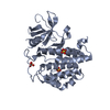
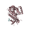
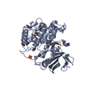
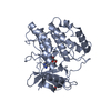
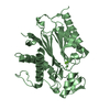
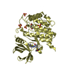
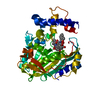
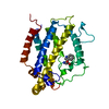
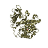
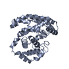
| Title | crystal structure of ck1d with PF670462 from P1 crystal form | ||||||
 Components Components | Casein kinase I isoform delta | ||||||
 Keywords Keywords | TRANSFERASE/TRANSFERASE INHIBITOR / ck1d / kinase / inhibitor / PF670462 / TRANSFERASE-TRANSFERASE INHIBITOR complex | ||||||
| Function / homology |  Function and homology information Function and homology informationpositive regulation of non-canonical Wnt signaling pathway / protein localization to Golgi apparatus / COPII vesicle coat assembly / tau-protein kinase / microtubule nucleation / protein localization to cilium / The CRY:PER:kinase complex represses transactivation by the BMAL:CLOCK (ARNTL:CLOCK) complex / midbrain dopaminergic neuron differentiation / non-motile cilium assembly / protein localization to centrosome ...positive regulation of non-canonical Wnt signaling pathway / protein localization to Golgi apparatus / COPII vesicle coat assembly / tau-protein kinase / microtubule nucleation / protein localization to cilium / The CRY:PER:kinase complex represses transactivation by the BMAL:CLOCK (ARNTL:CLOCK) complex / midbrain dopaminergic neuron differentiation / non-motile cilium assembly / protein localization to centrosome / COPII-mediated vesicle transport / Phosphorylation and nuclear translocation of the CRY:PER:kinase complex / tau-protein kinase activity / Golgi organization / Major pathway of rRNA processing in the nucleolus and cytosol / spindle assembly / endoplasmic reticulum-Golgi intermediate compartment membrane / Loss of Nlp from mitotic centrosomes / Loss of proteins required for interphase microtubule organization from the centrosome / Recruitment of mitotic centrosome proteins and complexes / Recruitment of NuMA to mitotic centrosomes / Anchoring of the basal body to the plasma membrane / AURKA Activation by TPX2 / spindle microtubule / circadian regulation of gene expression / sperm end piece / regulation of circadian rhythm / spindle / Wnt signaling pathway / endocytosis / Regulation of PLK1 Activity at G2/M Transition / positive regulation of canonical Wnt signaling pathway / positive regulation of proteasomal ubiquitin-dependent protein catabolic process / actin cytoskeleton / sperm principal piece / protein phosphorylation / protein kinase activity / non-specific serine/threonine protein kinase / cilium / ciliary basal body / cadherin binding / protein serine kinase activity / protein serine/threonine kinase activity / centrosome / perinuclear region of cytoplasm / Golgi apparatus / signal transduction / nucleoplasm / ATP binding / nucleus / plasma membrane / cytoplasm / cytosol Similarity search - Function | ||||||
| Biological species |  Homo sapiens (human) Homo sapiens (human) | ||||||
| Method |  X-RAY DIFFRACTION / SYNCHROTRON / MOLECULAR REPLACEMENT / Resolution: 2 Å X-RAY DIFFRACTION / SYNCHROTRON / MOLECULAR REPLACEMENT / Resolution: 2 Å | ||||||
 Authors Authors | Huang, X. | ||||||
 Citation Citation | Journal: J.Med.Chem. / Year: 2012 Title: Structural basis for the interaction between casein kinase 1 delta and a potent and selective inhibitor. Authors: Long, A. / Zhao, H. / Huang, X. | ||||||
| History |
|
- Structure visualization
| Structure viewer | Molecule: MolmilJmol/JSmol |
|---|
- Downloads & links
-Download
| PDBx/mmCIF format | 3uyt.cif.gz | 257.8 KB | Display | PDBx/mmCIF format |
|---|---|---|---|---|
| PDB format | pdb3uyt.ent.gz | 206.7 KB | Display | PDB format |
| PDBx/mmJSON format | 3uyt.json.gz | Tree view | PDBx/mmJSON format | |
| Others |  Other downloads Other downloads |
-Validation report
| Arichive directory | https://data.pdbj.org/pub/pdb/validation_reports/uy/3uytftp://data.pdbj.org/pub/pdb/validation_reports/uy/3uyt | HTTPS FTP |
|---|
-Related structure data


-Links
 PDBj
PDBj


- Assembly
| Deposited unit | 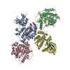
| ||||||||
|---|---|---|---|---|---|---|---|---|---|
| 1 | 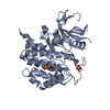
| ||||||||
| 2 | 
| ||||||||
| 3 | 
| ||||||||
| 4 | 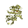
| ||||||||
| 5 | 
| ||||||||
| 6 | 
| ||||||||
| Unit cell |
|
-Components
| #1: Protein | Mass: 34350.660 Da / Num. of mol.: 4 Source method: isolated from a genetically manipulated source Source: (gene. exp.) Homo sapiens (human) / Gene: CSNK1D / Production host:   Spodoptera frugiperda (fall armyworm) Spodoptera frugiperda (fall armyworm)References: UniProt: P48730, non-specific serine/threonine protein kinase #2: Chemical | ChemComp-0CK /   Mass: 337.394 Da / Num. of mol.: 4 / Source method: obtained synthetically / Formula: C19H20FN5 Mass: 337.394 Da / Num. of mol.: 4 / Source method: obtained synthetically / Formula: C19H20FN5#3: Chemical | ChemComp-SO4 /   Mass: 96.063 Da / Num. of mol.: 7 / Source method: obtained synthetically / Formula: SO4 Mass: 96.063 Da / Num. of mol.: 7 / Source method: obtained synthetically / Formula: SO4#4: Water | ChemComp-HOH / |  Mass: 18.015 Da / Num. of mol.: 940 / Source method: isolated from a natural source / Formula: H2O Mass: 18.015 Da / Num. of mol.: 940 / Source method: isolated from a natural source / Formula: H2O |
|---|
-Experimental details
-Experiment
| Experiment | Method: X-RAY DIFFRACTION / Number of used crystals: 1 |
|---|
- Sample preparation
| Crystal | Density Matthews: 2.5 Å3/Da / Density % sol: 50.85 % |
|---|---|
| Crystal grow | Temperature: 277 K / Method: vapor diffusion, hanging drop / pH: 5 Details: 100 mM Citrate pH5.0, 0.2 or 0.4 M Na2SO4, 15-20% PEG3350, VAPOR DIFFUSION, HANGING DROP, temperature 277K |
-Data collection
| Diffraction | Mean temperature: 100 K |
|---|---|
| Diffraction source | Source: SYNCHROTRON / Site: APS  / Beamline: 21-ID-F / Wavelength: 0.9787 Å / Beamline: 21-ID-F / Wavelength: 0.9787 Å |
| Detector | Type: RAYONIX MX-225 / Detector: CCD / Date: Aug 1, 2011 |
| Radiation | Protocol: SINGLE WAVELENGTH / Monochromatic (M) / Laue (L): M / Scattering type: x-ray |
| Radiation wavelength | Wavelength: 0.9787 Å / Relative weight: 1 |
| Reflection | Resolution: 1.92→50 Å / Num. obs: 97538 / % possible obs: 97.6 % / Observed criterion σ(I): -3 / Rmerge(I) obs: 0.075 |
| Reflection shell | Resolution: 1.92→1.99 Å / Rmerge(I) obs: 0.534 / % possible all: 95.2 |
- Processing
| Software |
| ||||||||||||||||
|---|---|---|---|---|---|---|---|---|---|---|---|---|---|---|---|---|---|
| Refinement | Method to determine structure: MOLECULAR REPLACEMENT / Resolution: 2→50 Å / σ(F): 0 / Stereochemistry target values: Engh & Huber
| ||||||||||||||||
| Refinement step | Cycle: LAST / Resolution: 2→50 Å
|
