 Movie
Movie Controller

[English] 日本語
 Yorodumi
- PDB-3sc6: 2.65 Angstrom resolution crystal structure of dTDP-4-dehydrorhamn... -
+ Open data

- Basic information
| Entry | Database: PDB / ID: 3sc6 | ||||||
|---|---|---|---|---|---|---|---|
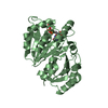
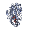
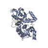
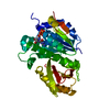
| Title | 2.65 Angstrom resolution crystal structure of dTDP-4-dehydrorhamnose reductase (rfbD) from Bacillus anthracis str. Ames in complex with NADP | ||||||
 Components Components | dTDP-4-dehydrorhamnose reductase | ||||||
 Keywords Keywords | OXIDOREDUCTASE / dTDP-4-dehydrorhamnose reductase / rfbD / structural genomics / infectious diseases / Bacillus anthracis str. Ames / rhamnose biosynthetic pathway / Center for Structural Genomics of Infectious Diseases / CSGID / Rossmann fold / Catalyzes formation of dTDP-4-dehydro-6-deoxy-L-mannose / NADPH and H+ from dTDP-6-deoxy-L-mannose and NADP+ | ||||||
| Function / homology |  Function and homology information Function and homology informationdTDP-4-dehydrorhamnose reductase / dTDP-4-dehydrorhamnose reductase activity / dTDP-rhamnose biosynthetic process / nucleotide binding / cytosol Similarity search - Function | ||||||
| Biological species |  | ||||||
| Method |  X-RAY DIFFRACTION / SYNCHROTRON / MOLECULAR REPLACEMENT / Resolution: 2.65 Å X-RAY DIFFRACTION / SYNCHROTRON / MOLECULAR REPLACEMENT / Resolution: 2.65 Å | ||||||
 Authors Authors | Halavaty, A.S. / Kuhn, M. / Shuvalova, L. / Minasov, G. / Peterson, S. / Anderson, W.F. / Center for Structural Genomics of Infectious Diseases (CSGID) | ||||||
 Citation Citation | Journal: Acta Crystallogr F Struct Biol Commun / Year: 2017 Title: Structure of the Bacillus anthracis dTDP-L-rhamnose-biosynthetic enzyme dTDP-4-dehydrorhamnose reductase (RfbD). Authors: Law, A. / Stergioulis, A. / Halavaty, A.S. / Minasov, G. / Anderson, W.F. / Kuhn, M.L. | ||||||
| History |
|
- Structure visualization
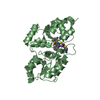
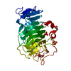
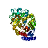
| Structure viewer | Molecule: MolmilJmol/JSmol |
|---|
- Downloads & links
-Download
| PDBx/mmCIF format | 3sc6.cif.gz | 689 KB | Display | PDBx/mmCIF format |
|---|---|---|---|---|
| PDB format | pdb3sc6.ent.gz | 580.2 KB | Display | PDB format |
| PDBx/mmJSON format | 3sc6.json.gz | Tree view | PDBx/mmJSON format | |
| Others |  Other downloads Other downloads |
-Validation report
| Arichive directory | https://data.pdbj.org/pub/pdb/validation_reports/sc/3sc6ftp://data.pdbj.org/pub/pdb/validation_reports/sc/3sc6 | HTTPS FTP |
|---|
-Related structure data
| Related structure data |  1vl0S S: Starting model for refinement |
|---|---|
| Similar structure data | |
| Other databases |

-Links
 PDBj
PDBj- Assembly
| Deposited unit | 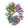
| ||||||||
|---|---|---|---|---|---|---|---|---|---|
| 1 | 
| ||||||||
| 2 | 
| ||||||||
| 3 | 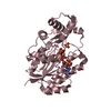
| ||||||||
| 4 | 
| ||||||||
| 5 | 
| ||||||||
| 6 | 
| ||||||||
| Unit cell |
|
-Components
| #1: Protein | Mass: 32721.137 Da / Num. of mol.: 6 Source method: isolated from a genetically manipulated source Source: (gene. exp.) References: UniProt: Q81TN9, UniProt: A0A6L7H7R4*PLUS, dTDP-4-dehydrorhamnose reductase #2: Chemical | ChemComp-NAP /   Mass: 743.405 Da / Num. of mol.: 6 / Source method: obtained synthetically / Formula: C21H28N7O17P3 Mass: 743.405 Da / Num. of mol.: 6 / Source method: obtained synthetically / Formula: C21H28N7O17P3#3: Chemical | ChemComp-SO4 /   Mass: 96.063 Da / Num. of mol.: 17 / Source method: obtained synthetically / Formula: SO4 Mass: 96.063 Da / Num. of mol.: 17 / Source method: obtained synthetically / Formula: SO4#4: Water | ChemComp-HOH / |  Mass: 18.015 Da / Num. of mol.: 240 / Source method: isolated from a natural source / Formula: H2O Mass: 18.015 Da / Num. of mol.: 240 / Source method: isolated from a natural source / Formula: H2O |
|---|
-Experimental details
-Experiment
| Experiment | Method: X-RAY DIFFRACTION / Number of used crystals: 1 |
|---|
- Sample preparation
| Crystal | Density Matthews: 3.32 Å3/Da / Density % sol: 62.99 % |
|---|---|
| Crystal grow | Temperature: 297 K / Method: vapor diffusion, sitting drop / pH: 8.5 Details: Protein: 7.5 mg/mL in buffer containing 10 mM Tris HCl pH 8.3, 500 mM NaCl, and 5 mM BME, 5 mM MgCl2, 1 mM NADP+, and 10% glycerol.Crystallization conditions: 0.2 M lithium sulfate, 0.1 M ...Details: Protein: 7.5 mg/mL in buffer containing 10 mM Tris HCl pH 8.3, 500 mM NaCl, and 5 mM BME, 5 mM MgCl2, 1 mM NADP+, and 10% glycerol.Crystallization conditions: 0.2 M lithium sulfate, 0.1 M Tris pH 8.5, and 25% w/v PEG 5000 MME |
-Data collection
| Diffraction | Mean temperature: 100 K |
|---|---|
| Diffraction source | Source: SYNCHROTRON / Site: APS  / Beamline: 21-ID-D / Wavelength: 0.97856 Å / Beamline: 21-ID-D / Wavelength: 0.97856 Å |
| Detector | Type: MARMOSAIC 300 mm CCD / Detector: CCD / Date: Mar 3, 2011 / Details: Mirror |
| Radiation | Monochromator: Si(111) Channel / Protocol: SINGLE WAVELENGTH / Monochromatic (M) / Laue (L): M / Scattering type: x-ray |
| Radiation wavelength | Wavelength: 0.97856 Å / Relative weight: 1 |
| Reflection | Resolution: 2.65→30 Å / Num. all: 75200 / Num. obs: 75200 / % possible obs: 100 % / Observed criterion σ(I): -3 / Redundancy: 3.8 % / Biso Wilson estimate: 67.1 Å2 / Rmerge(I) obs: 0.102 / Net I/σ(I): 11.49 |
| Reflection shell | Resolution: 2.65→2.7 Å / Redundancy: 3.8 % / Rmerge(I) obs: 0.615 / Mean I/σ(I) obs: 2.35 / Num. unique all: 3724 / % possible all: 100 |
- Processing
| Software |
| |||||||||||||||||||||||||||||||||||||||||||||||||||||||||||||||||||||||||||||||||||||||||||||||||||||||||||||||||||||||||||||||||||||||||||||||||||||||||||||||||||||||||||||||||||||||||||||||||||||||||||||||||||||||||||||||||||||||||||||||||||||||||||||||||||||||||||||||||||||||||||||||||||||||||||||||||||||||||||||||||||||
|---|---|---|---|---|---|---|---|---|---|---|---|---|---|---|---|---|---|---|---|---|---|---|---|---|---|---|---|---|---|---|---|---|---|---|---|---|---|---|---|---|---|---|---|---|---|---|---|---|---|---|---|---|---|---|---|---|---|---|---|---|---|---|---|---|---|---|---|---|---|---|---|---|---|---|---|---|---|---|---|---|---|---|---|---|---|---|---|---|---|---|---|---|---|---|---|---|---|---|---|---|---|---|---|---|---|---|---|---|---|---|---|---|---|---|---|---|---|---|---|---|---|---|---|---|---|---|---|---|---|---|---|---|---|---|---|---|---|---|---|---|---|---|---|---|---|---|---|---|---|---|---|---|---|---|---|---|---|---|---|---|---|---|---|---|---|---|---|---|---|---|---|---|---|---|---|---|---|---|---|---|---|---|---|---|---|---|---|---|---|---|---|---|---|---|---|---|---|---|---|---|---|---|---|---|---|---|---|---|---|---|---|---|---|---|---|---|---|---|---|---|---|---|---|---|---|---|---|---|---|---|---|---|---|---|---|---|---|---|---|---|---|---|---|---|---|---|---|---|---|---|---|---|---|---|---|---|---|---|---|---|---|---|---|---|---|---|---|---|---|---|---|---|---|---|---|---|---|---|---|---|---|---|---|---|---|---|---|---|---|---|---|---|---|---|---|---|---|---|---|---|---|---|---|---|---|---|---|---|---|---|---|---|---|---|---|---|---|---|---|---|---|---|---|---|---|---|
| Refinement | Method to determine structure: MOLECULAR REPLACEMENT Starting model: 1VL0 Resolution: 2.65→29.9 Å / Cor.coef. Fo:Fc: 0.943 / Cor.coef. Fo:Fc free: 0.922 / SU B: 26.342 / SU ML: 0.252 / Isotropic thermal model: isotropic / Cross valid method: THROUGHOUT / ESU R Free: 0.313 / Stereochemistry target values: MAXIMUM LIKELIHOOD / Details: HYDROGENS HAVE BEEN ADDED IN THE RIDING POSITIONS
| |||||||||||||||||||||||||||||||||||||||||||||||||||||||||||||||||||||||||||||||||||||||||||||||||||||||||||||||||||||||||||||||||||||||||||||||||||||||||||||||||||||||||||||||||||||||||||||||||||||||||||||||||||||||||||||||||||||||||||||||||||||||||||||||||||||||||||||||||||||||||||||||||||||||||||||||||||||||||||||||||||||
| Solvent computation | Ion probe radii: 0.8 Å / Shrinkage radii: 0.8 Å / VDW probe radii: 1.2 Å / Solvent model: BABINET MODEL WITH MASK | |||||||||||||||||||||||||||||||||||||||||||||||||||||||||||||||||||||||||||||||||||||||||||||||||||||||||||||||||||||||||||||||||||||||||||||||||||||||||||||||||||||||||||||||||||||||||||||||||||||||||||||||||||||||||||||||||||||||||||||||||||||||||||||||||||||||||||||||||||||||||||||||||||||||||||||||||||||||||||||||||||||
| Displacement parameters | Biso mean: 56.691 Å2
| |||||||||||||||||||||||||||||||||||||||||||||||||||||||||||||||||||||||||||||||||||||||||||||||||||||||||||||||||||||||||||||||||||||||||||||||||||||||||||||||||||||||||||||||||||||||||||||||||||||||||||||||||||||||||||||||||||||||||||||||||||||||||||||||||||||||||||||||||||||||||||||||||||||||||||||||||||||||||||||||||||||
| Refinement step | Cycle: LAST / Resolution: 2.65→29.9 Å
| |||||||||||||||||||||||||||||||||||||||||||||||||||||||||||||||||||||||||||||||||||||||||||||||||||||||||||||||||||||||||||||||||||||||||||||||||||||||||||||||||||||||||||||||||||||||||||||||||||||||||||||||||||||||||||||||||||||||||||||||||||||||||||||||||||||||||||||||||||||||||||||||||||||||||||||||||||||||||||||||||||||
| Refine LS restraints |
| |||||||||||||||||||||||||||||||||||||||||||||||||||||||||||||||||||||||||||||||||||||||||||||||||||||||||||||||||||||||||||||||||||||||||||||||||||||||||||||||||||||||||||||||||||||||||||||||||||||||||||||||||||||||||||||||||||||||||||||||||||||||||||||||||||||||||||||||||||||||||||||||||||||||||||||||||||||||||||||||||||||
| LS refinement shell | Resolution: 2.65→2.718 Å / Total num. of bins used: 20
| |||||||||||||||||||||||||||||||||||||||||||||||||||||||||||||||||||||||||||||||||||||||||||||||||||||||||||||||||||||||||||||||||||||||||||||||||||||||||||||||||||||||||||||||||||||||||||||||||||||||||||||||||||||||||||||||||||||||||||||||||||||||||||||||||||||||||||||||||||||||||||||||||||||||||||||||||||||||||||||||||||||
| Refinement TLS params. | Method: refined / Refine-ID: X-RAY DIFFRACTION
| |||||||||||||||||||||||||||||||||||||||||||||||||||||||||||||||||||||||||||||||||||||||||||||||||||||||||||||||||||||||||||||||||||||||||||||||||||||||||||||||||||||||||||||||||||||||||||||||||||||||||||||||||||||||||||||||||||||||||||||||||||||||||||||||||||||||||||||||||||||||||||||||||||||||||||||||||||||||||||||||||||||
| Refinement TLS group |
|
