 Movie
Movie Controller

[English] 日本語
 Yorodumi
- PDB-3fkg: Crystal Structure Analysis of Fungal Versatile Peroxidase from Pl... -
+ Open data

- Basic information
| Entry | Database: PDB / ID: 3fkg | ||||||
|---|---|---|---|---|---|---|---|
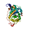
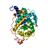
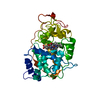
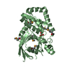
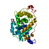
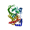
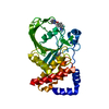
| Title | Crystal Structure Analysis of Fungal Versatile Peroxidase from Pleurotus eryngii | ||||||
 Components Components | Versatile peroxidase VPL2 | ||||||
 Keywords Keywords | OXIDOREDUCTASE / ALLELIC VARIANT / AROMATIC-SUBSTRATE BINDING / CLASS II (FUNGAL)PEROXIDASES / protoporphyrin IX / ELECTRON TRANSFER / LIGNIN PEROXIDASE / LIGNIN DEGRADATION / MANGANESE PEROXIDASE / MN-INDEPENDENT OXIDATION PHENOLIC NON-PHENOLIC AROMATICS / MNII OXIDATION / PEROXIDASE / POLYVALENT PEROXIDASE / Heme / Hydrogen peroxide / Iron / Manganese / Metal-binding / Secreted / Zymogen | ||||||
| Function / homology |  Function and homology information Function and homology informationversatile peroxidase / versatile peroxidase activity / manganese peroxidase activity / lignin catabolic process / hydrogen peroxide catabolic process / response to reactive oxygen species / cellular response to oxidative stress / heme binding / extracellular region / metal ion binding Similarity search - Function | ||||||
| Biological species |  Pleurotus eryngii (fungus) Pleurotus eryngii (fungus) | ||||||
| Method |  X-RAY DIFFRACTION / SYNCHROTRON / MOLECULAR REPLACEMENT / Resolution: 1.81 Å X-RAY DIFFRACTION / SYNCHROTRON / MOLECULAR REPLACEMENT / Resolution: 1.81 Å | ||||||
 Authors Authors | Piontek, K. / Martinez, A.T. / Choinowski, T. / Plattner, D.A. | ||||||
 Citation Citation | Journal: To be Published Title: Structural and Site-directed Mutagenesis Study of Versatile Peroxidase Oxidizing both Mn(II) and Aromatic Substrates Authors: Piontek, K. / Choinowski, T. / Perez-Boada, M. / Ruiz-Duenas, F.J. / Martinez, M.J. / Plattner, D.A. / Martinez, A.T. | ||||||
| History |
|
- Structure visualization
| Structure viewer | Molecule: MolmilJmol/JSmol |
|---|
- Downloads & links
-Download
| PDBx/mmCIF format | 3fkg.cif.gz | 88 KB | Display | PDBx/mmCIF format |
|---|---|---|---|---|
| PDB format | pdb3fkg.ent.gz | 65.6 KB | Display | PDB format |
| PDBx/mmJSON format | 3fkg.json.gz | Tree view | PDBx/mmJSON format | |
| Others |  Other downloads Other downloads |
-Validation report
| Arichive directory | https://data.pdbj.org/pub/pdb/validation_reports/fk/3fkgftp://data.pdbj.org/pub/pdb/validation_reports/fk/3fkg | HTTPS FTP |
|---|
-Related structure data
| Related structure data |  3fjwC  3fm1C  3fm4C  3fm6C  3fmuC 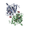 1qpaS C: citing same article ( S: Starting model for refinement |
|---|---|
| Similar structure data |


-Links
 PDBj
PDBj


- Assembly
| Deposited unit | 
| ||||||||
|---|---|---|---|---|---|---|---|---|---|
| 1 |
| ||||||||
| Unit cell |
|
-Components
-Protein , 1 types, 1 molecules A
| #1: Protein | Mass: 34663.734 Da / Num. of mol.: 1 Source method: isolated from a genetically manipulated source Source: (gene. exp.) Pleurotus eryngii (fungus) / Strain: IJFM, A169 / Gene: vpl2 / Plasmid: PFLAG1-VPL2 / Production host:  |
|---|
-Non-polymers , 6 types, 365 molecules 


| #2: Chemical | ChemComp-HEM /  Mass: 616.487 Da / Num. of mol.: 1 / Source method: obtained synthetically / Formula: C34H32FeN4O4 Mass: 616.487 Da / Num. of mol.: 1 / Source method: obtained synthetically / Formula: C34H32FeN4O4 | ||||||||
|---|---|---|---|---|---|---|---|---|---|
| #3: Chemical |  Mass: 40.078 Da / Num. of mol.: 2 / Source method: obtained synthetically / Formula: Ca Mass: 40.078 Da / Num. of mol.: 2 / Source method: obtained synthetically / Formula: Ca#4: Chemical | ChemComp-ZN /  Mass: 65.409 Da / Num. of mol.: 7 / Source method: obtained synthetically / Formula: Zn Mass: 65.409 Da / Num. of mol.: 7 / Source method: obtained synthetically / Formula: Zn#5: Chemical |  Mass: 55.845 Da / Num. of mol.: 3 / Source method: obtained synthetically / Formula: Fe Mass: 55.845 Da / Num. of mol.: 3 / Source method: obtained synthetically / Formula: Fe#6: Chemical |  Mass: 136.989 Da / Num. of mol.: 2 / Source method: obtained synthetically / Formula: C2H6AsO2 Mass: 136.989 Da / Num. of mol.: 2 / Source method: obtained synthetically / Formula: C2H6AsO2#7: Water | ChemComp-HOH / | Mass: 18.015 Da / Num. of mol.: 350 / Source method: isolated from a natural source / Formula: H2O |
-Details
| Has protein modification | Y |
|---|---|
| Sequence details | THERE ARE CONFLICTS BETWEEN THE REPORTED SEQUENCE AND THE DATABASE REFERENCE SEQUENCE. THIS CAN BE ...THERE ARE CONFLICTS BETWEEN THE REPORTED SEQUENCE AND THE DATABASE REFERENCE SEQUENCE. THIS CAN BE CALLED AS SPONTANEIT |
-Experimental details
-Experiment
| Experiment | Method: X-RAY DIFFRACTION / Number of used crystals: 1 |
|---|
- Sample preparation
| Crystal | Density Matthews: 2.78 Å3/Da / Density % sol: 55.81 % |
|---|---|
| Crystal grow | Temperature: 298 K / Method: vapor diffusion, hanging drop / pH: 6.5 Details: 9.0mg/ml protein in 10mM Na-tartrate pH 5.5, 17% PEG 10000, 200mM Zn-acetate, 100mM Na-cacodylate pH 6.5, VAPOR DIFFUSION, HANGING DROP, temperature 298K |
-Data collection
| Diffraction | Mean temperature: 100 K |
|---|---|
| Diffraction source | Source: SYNCHROTRON / Site: EMBL/DESY, HAMBURG  / Beamline: X11 / Wavelength: 0.8065 Å / Beamline: X11 / Wavelength: 0.8065 Å |
| Detector | Type: MARMOSAIC 325 mm CCD / Detector: CCD / Date: Nov 14, 2001 |
| Radiation | Protocol: SINGLE WAVELENGTH / Monochromatic (M) / Laue (L): M / Scattering type: x-ray |
| Radiation wavelength | Wavelength: 0.8065 Å / Relative weight: 1 |
| Reflection | Resolution: 1.81→40 Å / Num. all: 34670 / Num. obs: 33318 / % possible obs: 96.1 % / Observed criterion σ(F): 1 / Observed criterion σ(I): 1 / Redundancy: 2.4 % / Biso Wilson estimate: 20.3 Å2 / Rsym value: 0.056 / Net I/σ(I): 11.9 |
| Reflection shell | Resolution: 1.81→1.84 Å / Mean I/σ(I) obs: 1.1 / Rsym value: 0.392 / % possible all: 98.6 |
- Processing
| Software |
| ||||||||||||||||||||||||||||||||||||||||||||||||||||||||||||||||||||||||||||||||||||||||||||||||||||
|---|---|---|---|---|---|---|---|---|---|---|---|---|---|---|---|---|---|---|---|---|---|---|---|---|---|---|---|---|---|---|---|---|---|---|---|---|---|---|---|---|---|---|---|---|---|---|---|---|---|---|---|---|---|---|---|---|---|---|---|---|---|---|---|---|---|---|---|---|---|---|---|---|---|---|---|---|---|---|---|---|---|---|---|---|---|---|---|---|---|---|---|---|---|---|---|---|---|---|---|---|---|
| Refinement | Method to determine structure: MOLECULAR REPLACEMENT Starting model: PDB ENTRY 1QPA Resolution: 1.81→32.9 Å / Cor.coef. Fo:Fc: 0.968 / Cor.coef. Fo:Fc free: 0.948 / SU B: 1.617 / SU ML: 0.051 / Cross valid method: THROUGHOUT / σ(F): 0 / ESU R: 0.106 / ESU R Free: 0.112 / Stereochemistry target values: MAXIMUM LIKELIHOOD
| ||||||||||||||||||||||||||||||||||||||||||||||||||||||||||||||||||||||||||||||||||||||||||||||||||||
| Solvent computation | Ion probe radii: 0.8 Å / Shrinkage radii: 0.8 Å / VDW probe radii: 1.2 Å / Solvent model: BABINET MODEL WITH MASK | ||||||||||||||||||||||||||||||||||||||||||||||||||||||||||||||||||||||||||||||||||||||||||||||||||||
| Displacement parameters | Biso mean: 25.889 Å2
| ||||||||||||||||||||||||||||||||||||||||||||||||||||||||||||||||||||||||||||||||||||||||||||||||||||
| Refinement step | Cycle: LAST / Resolution: 1.81→32.9 Å
| ||||||||||||||||||||||||||||||||||||||||||||||||||||||||||||||||||||||||||||||||||||||||||||||||||||
| Refine LS restraints |
| ||||||||||||||||||||||||||||||||||||||||||||||||||||||||||||||||||||||||||||||||||||||||||||||||||||
| LS refinement shell | Resolution: 1.81→1.857 Å / Total num. of bins used: 20
|
