[English] 日本語
Yorodumi
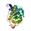
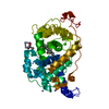
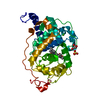
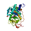
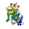
- PDB-5abo: CRYSTAL STRUCTURE ANALYSIS OF FUNGAL VERSATILE PEROXIDASE FROM PL... -

+
Open data


ID or keywords:

Loading...

-
Basic information

Entry
Database: PDB / ID: 5abo
TitleCRYSTAL STRUCTURE ANALYSIS OF FUNGAL VERSATILE PEROXIDASE FROM PLEUROTUS ERYNGII. MUTANT VPi-br. MUTATED RESIDUES T2K, D69S, T70D, S86E, A131K, D146T, Q202L, Q219K, H232E, Q239R, L288R, S301K, A308R, A309K AND A314R.
ComponentsVERSATILE PEROXIDASE VPL2
KeywordsOXIDOREDUCTASE / CLASS II (FUNGAL) PEROXIDASES / PROTOPORPHYRIN IX / ELECTRON TRANSFER / LIGNIN PEROXIDASE / LIGNIN DEGRADATION / MANGANESE PEROXIDASE / MN-INDEPENDENT OXIDATION PHENOLIC NON-PHENOLIC AROMATICS / MNII OXIDATION / PEROXIDASE / POLYVALENT PEROXIDASE / HEME / HYDROGEN PEROXIDE / IRON / MANGANESE / METAL-BINDING / SECRETED / ZYMOGEN
Function / homology
Function and homology information


versatile peroxidase / reactive-black-5:hydrogen-peroxide oxidoreductase activity / manganese peroxidase activity / lignin catabolic process / response to reactive oxygen species / hydrogen peroxide catabolic process / cellular response to oxidative stress / heme binding / extracellular region / metal ion binding
Similarity search - Function
Fungal ligninase / Fungal ligninase, C-terminal / Fungal peroxidase extension region / Heme-binding peroxidase Ccp1-like / Peroxidase; domain 2 / Peroxidase, domain 2 / Peroxidase; domain 1 - #10 / Peroxidase; domain 1 / Peroxidases heam-ligand binding site / Peroxidase, active site ...Fungal ligninase / Fungal ligninase, C-terminal / Fungal peroxidase extension region / Heme-binding peroxidase Ccp1-like / Peroxidase; domain 2 / Peroxidase, domain 2 / Peroxidase; domain 1 - #10 / Peroxidase; domain 1 / Peroxidases heam-ligand binding site / Peroxidase, active site / Peroxidases active site signature. / Plant heme peroxidase family profile. / Haem peroxidase / Peroxidase / Peroxidases proximal heme-ligand signature. / Haem peroxidase superfamily / Orthogonal Bundle / Mainly Alpha
Similarity search - Domain/homology
PROTOPORPHYRIN IX CONTAINING FE / Versatile peroxidase VPL2
Similarity search - Component
Biological speciesPLEUROTUS ERYNGII (fungus)
MethodX-RAY DIFFRACTION / SYNCHROTRON / MOLECULAR REPLACEMENT / Resolution: 1.095 Å
AuthorsMedrano, F.J. / Romero, A.
CitationJournal: Plos One / Year: 2015
Title: Improving the Ph-Stability of Versatile Peroxidase by Comparative Structural Analysis with a Naturally-Stable Manganese Peroxidase.
Authors: Saez-Jimenez, V. / Fernendez-Fueyo, E. / Medrano, F.J. / Romero, A. / Martinez, A.T. / Ruiz-Duenas, F.J.
History
DepositionAug 7, 2015Deposition site: PDBE / Processing site: PDBE
Revision 1.0Nov 4, 2015Provider: repository / Type: Initial release
Revision 1.1Jan 10, 2024Group: Data collection / Database references ...Data collection / Database references / Derived calculations / Other / Refinement description
Category: chem_comp_atom / chem_comp_bond ...chem_comp_atom / chem_comp_bond / database_2 / pdbx_database_status / pdbx_initial_refinement_model / pdbx_struct_conn_angle / struct_conn / struct_site
Item: _database_2.pdbx_DOI / _database_2.pdbx_database_accession ..._database_2.pdbx_DOI / _database_2.pdbx_database_accession / _pdbx_database_status.status_code_sf / _pdbx_struct_conn_angle.ptnr1_auth_comp_id / _pdbx_struct_conn_angle.ptnr1_auth_seq_id / _pdbx_struct_conn_angle.ptnr1_label_asym_id / _pdbx_struct_conn_angle.ptnr1_label_atom_id / _pdbx_struct_conn_angle.ptnr1_label_comp_id / _pdbx_struct_conn_angle.ptnr1_label_seq_id / _pdbx_struct_conn_angle.ptnr2_auth_comp_id / _pdbx_struct_conn_angle.ptnr2_auth_seq_id / _pdbx_struct_conn_angle.ptnr2_label_asym_id / _pdbx_struct_conn_angle.ptnr2_label_atom_id / _pdbx_struct_conn_angle.ptnr2_label_comp_id / _pdbx_struct_conn_angle.ptnr3_auth_comp_id / _pdbx_struct_conn_angle.ptnr3_auth_seq_id / _pdbx_struct_conn_angle.ptnr3_label_asym_id / _pdbx_struct_conn_angle.ptnr3_label_atom_id / _pdbx_struct_conn_angle.ptnr3_label_comp_id / _pdbx_struct_conn_angle.ptnr3_label_seq_id / _pdbx_struct_conn_angle.value / _struct_conn.pdbx_dist_value / _struct_conn.ptnr1_auth_comp_id / _struct_conn.ptnr1_auth_seq_id / _struct_conn.ptnr1_label_asym_id / _struct_conn.ptnr1_label_atom_id / _struct_conn.ptnr1_label_comp_id / _struct_conn.ptnr1_label_seq_id / _struct_conn.ptnr2_auth_comp_id / _struct_conn.ptnr2_auth_seq_id / _struct_conn.ptnr2_label_asym_id / _struct_conn.ptnr2_label_atom_id / _struct_conn.ptnr2_label_comp_id / _struct_conn.ptnr2_label_seq_id / _struct_site.pdbx_auth_asym_id / _struct_site.pdbx_auth_comp_id / _struct_site.pdbx_auth_seq_id

-
Structure visualization

Structure viewerMolecule:
MolmilJmol/JSmol

Downloads & links

-
Assembly

Deposited unit
A: VERSATILE PEROXIDASE VPL2
hetero molecules


Theoretical massNumber of molelcules
Total (without water)35,7254
Polymers35,0281
Non-polymers6973
Water8,089449
1


  • Idetical with deposited unit
  • defined by author&software
TypeNameSymmetry operationNumber
identity operation1_555x,y,z1
MethodPISA
Unit cell
Length a, b, c (Å)54.820, 64.220, 95.410
Angle α, β, γ (deg.)90.00, 90.00, 90.00
Int Tables number19
Space group name H-MP212121

-
Components

#1: Protein VERSATILE PEROXIDASE VPL2 / VERSATILE LIQUID PHASE PEROXIDASE 2 / VERSATILE PEROXIDASE


Mass: 35028.492 Da / Num. of mol.: 1 / Fragment: UNP RESIDUES 33-361 / Mutation: YES
Source method: isolated from a genetically manipulated source
Source: (gene. exp.) PLEUROTUS ERYNGII (fungus) / Strain: IJFM, A169 / Production host: ESCHERICHIA COLI (E. coli) / Strain (production host): W3110 / References: UniProt: O94753, versatile peroxidase
#2: Chemical ChemComp-CA / CALCIUM ION


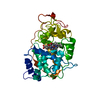
Mass: 40.078 Da / Num. of mol.: 2 / Source method: obtained synthetically / Formula: Ca
#3: Chemical ChemComp-HEM / PROTOPORPHYRIN IX CONTAINING FE / HEME


Mass: 616.487 Da / Num. of mol.: 1 / Source method: obtained synthetically / Formula: C34H32FeN4O4
#4: Water ChemComp-HOH / water


Mass: 18.015 Da / Num. of mol.: 449 / Source method: isolated from a natural source / Formula: H2O

-
Experimental details

-
Experiment

ExperimentMethod: X-RAY DIFFRACTION / Number of used crystals: 1

-
Sample preparation

CrystalDensity Matthews: 2.36 Å3/Da / Density % sol: 47.96 % / Description: NONE
Crystal growDetails: 10 % PEG 4000, 20% 2-PROPANOL

-
Data collection

DiffractionMean temperature: 100 K
Diffraction sourceSource: SYNCHROTRON / Site: SOLEIL / Beamline: PROXIMA 1 / Wavelength: 1
DetectorType: DECTRIS PILATUS 6M / Detector: PIXEL / Date: Nov 6, 2014
RadiationProtocol: SINGLE WAVELENGTH / Monochromatic (M) / Laue (L): M / Scattering type: x-ray
Radiation wavelengthWavelength: 1 Å / Relative weight: 1
ReflectionResolution: 1.1→50 Å / Num. obs: 135701 / % possible obs: 97.9 % / Observed criterion σ(I): 0 / Redundancy: 9.8 % / Biso Wilson estimate: 11.98 Å2 / Rmerge(I) obs: 0.07 / Net I/σ(I): 15.3
Reflection shellResolution: 1.1→1.16 Å / Redundancy: 6 % / Rmerge(I) obs: 1.09 / Mean I/σ(I) obs: 1.2 / % possible all: 86.7

-
Processing

Software
NameVersionClassification
PHENIX(PHENIX.REFINE)refinement
XDSdata reduction
XDSdata scaling
PHASERphasing
RefinementMethod to determine structure: MOLECULAR REPLACEMENT
Starting model: PDB ENTRY 3FMU
Resolution: 1.095→27.41 Å / SU ML: 0.1 / σ(F): 1.34 / Phase error: 14.06 / Stereochemistry target values: ML
RfactorNum. reflection% reflection
Rfree0.1441 2000 1.5 %
Rwork0.1335 --
obs0.1337 135667 97.85 %
Solvent computationShrinkage radii: 0.9 Å / VDW probe radii: 1.11 Å / Solvent model: FLAT BULK SOLVENT MODEL
Refinement stepCycle: LAST / Resolution: 1.095→27.41 Å
ProteinNucleic acidLigandSolventTotal
Num. atoms2358 0 45 449 2852
Refine LS restraints
Refine-IDTypeDev idealNumber
X-RAY DIFFRACTIONf_bond_d0.012469
X-RAY DIFFRACTIONf_angle_d1.4543365
X-RAY DIFFRACTIONf_dihedral_angle_d14.191890
X-RAY DIFFRACTIONf_chiral_restr0.088366
X-RAY DIFFRACTIONf_plane_restr0.009449
LS refinement shell
Resolution (Å)Rfactor RfreeNum. reflection RfreeRfactor RworkNum. reflection RworkRefine-ID% reflection obs (%)
1.0953-1.12270.29171090.29237333X-RAY DIFFRACTION76
1.1227-1.1530.2651370.23199059X-RAY DIFFRACTION94
1.153-1.1870.20311440.1889638X-RAY DIFFRACTION100
1.187-1.22530.18511450.16629694X-RAY DIFFRACTION100
1.2253-1.26910.16881440.15419644X-RAY DIFFRACTION100
1.2691-1.31990.15251450.13489702X-RAY DIFFRACTION100
1.3199-1.37990.14781450.12499678X-RAY DIFFRACTION100
1.3799-1.45270.13751450.11269721X-RAY DIFFRACTION100
1.4527-1.54370.12661460.10489722X-RAY DIFFRACTION100
1.5437-1.66290.1211460.10239757X-RAY DIFFRACTION100
1.6629-1.83020.10551460.11479795X-RAY DIFFRACTION100
1.8302-2.09490.14841470.12819799X-RAY DIFFRACTION100
2.0949-2.63910.13921480.13139931X-RAY DIFFRACTION100
2.6391-27.41860.14081530.136210194X-RAY DIFFRACTION100

+
About Yorodumi

-
News

-
Feb 9, 2022. New format data for meta-information of EMDB entries

New format data for meta-information of EMDB entries

  • Version 3 of the EMDB header file is now the official format.
  • The previous official version 1.9 will be removed from the archive.

Related info.:EMDB header

External links:wwPDB to switch to version 3 of the EMDB data model

-
Aug 12, 2020. Covid-19 info

Covid-19 info

URL: https://pdbjlvh1.pdbj.org/emnavi/covid19.php

New page: Covid-19 featured information page in EM Navigator.

Related info.:Covid-19 info / Mar 5, 2020. Novel coronavirus structure data

+
Mar 5, 2020. Novel coronavirus structure data

Novel coronavirus structure data

Related info.:Yorodumi Speices / Aug 12, 2020. Covid-19 info

External links:COVID-19 featured content - PDBj / Molecule of the Month (242):Coronavirus Proteases

+
Jan 31, 2019. EMDB accession codes are about to change! (news from PDBe EMDB page)

EMDB accession codes are about to change! (news from PDBe EMDB page)

  • The allocation of 4 digits for EMDB accession codes will soon come to an end. Whilst these codes will remain in use, new EMDB accession codes will include an additional digit and will expand incrementally as the available range of codes is exhausted. The current 4-digit format prefixed with “EMD-” (i.e. EMD-XXXX) will advance to a 5-digit format (i.e. EMD-XXXXX), and so on. It is currently estimated that the 4-digit codes will be depleted around Spring 2019, at which point the 5-digit format will come into force.
  • The EM Navigator/Yorodumi systems omit the EMD- prefix.

Related info.:Q: What is EMD? / ID/Accession-code notation in Yorodumi/EM Navigator

External links:EMDB Accession Codes are Changing Soon! / Contact to PDBj

+
Jul 12, 2017. Major update of PDB

Major update of PDB

  • wwPDB released updated PDB data conforming to the new PDBx/mmCIF dictionary.
  • This is a major update changing the version number from 4 to 5, and with Remediation, in which all the entries are updated.
  • In this update, many items about electron microscopy experimental information are reorganized (e.g. em_software).
  • Now, EM Navigator and Yorodumi are based on the updated data.

External links:wwPDB Remediation / Enriched Model Files Conforming to OneDep Data Standards Now Available in the PDB FTP Archive

-
Yorodumi

Thousand views of thousand structures

  • Yorodumi is a browser for structure data from EMDB, PDB, SASBDB, etc.
  • This page is also the successor to EM Navigator detail page, and also detail information page/front-end page for Omokage search.
  • The word "yorodu" (or yorozu) is an old Japanese word meaning "ten thousand". "mi" (miru) is to see.

Related info.:EMDB / PDB / SASBDB / Comparison of 3 databanks / Yorodumi Search / Aug 31, 2016. New EM Navigator & Yorodumi / Yorodumi Papers / Jmol/JSmol / Function and homology information / Changes in new EM Navigator and Yorodumi

Read more