 Movie
Movie Controller

[English] 日本語
 Yorodumi
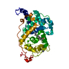
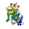
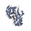
- PDB-4g05: The crystal structures of several mutants of Pleurotus eryngii ve... -
+ Open data

- Basic information
| Entry | Database: PDB / ID: 4g05 | ||||||
|---|---|---|---|---|---|---|---|
| Title | The crystal structures of several mutants of Pleurotus eryngii versatile peroxidase | ||||||
 Components Components | Versatile peroxidase VPL2 | ||||||
 Keywords Keywords | OXIDOREDUCTASE / LIGNIN PEROXIDASE / LIGNIN DEGRADATION / AROMATIC-SUBSTRATE BINDING | ||||||
| Function / homology |  Function and homology information Function and homology informationversatile peroxidase / versatile peroxidase activity / manganese peroxidase activity / lignin catabolic process / hydrogen peroxide catabolic process / response to reactive oxygen species / cellular response to oxidative stress / heme binding / extracellular region / metal ion binding Similarity search - Function | ||||||
| Biological species |  Pleurotus eryngii (fungus) Pleurotus eryngii (fungus) | ||||||
| Method |  X-RAY DIFFRACTION / SYNCHROTRON / MOLECULAR REPLACEMENT / Resolution: 2.35 Å X-RAY DIFFRACTION / SYNCHROTRON / MOLECULAR REPLACEMENT / Resolution: 2.35 Å | ||||||
 Authors Authors | Mate, M.J. / Romero, A. / Ruiz-Duenas, F.J. / Martinez, A.T. | ||||||
 Citation Citation | Journal: J.Biol.Chem. / Year: 2012 Title: Two Oxidation Sites for Low Redox Potential Substrates: A DIRECTED MUTAGENESIS, KINETIC, AND CRYSTALLOGRAPHIC STUDY ON PLEUROTUS ERYNGII VERSATILE PEROXIDASE. Authors: Morales, M. / Mate, M.J. / Romero, A. / Martinez, M.J. / Martinez, A.T. / Ruiz-Duenas, F.J. #1: Journal: Biochemistry / Year: 2008Title: Site-directed mutagenesis of the catalytic tryptophan environment in pleurotus eryngii versatile peroxidase Authors: Ruiz-Duenas, F.J. / Morales, M. / Mate, M.J. / Romero, A. / Martinez, M.J. / Smith, A. / Martinez, A.T. | ||||||
| History |
|
- Structure visualization
| Structure viewer | Molecule: MolmilJmol/JSmol |
|---|
- Downloads & links
-Download
| PDBx/mmCIF format | 4g05.cif.gz | 77.9 KB | Display | PDBx/mmCIF format |
|---|---|---|---|---|
| PDB format | pdb4g05.ent.gz | 56.8 KB | Display | PDB format |
| PDBx/mmJSON format | 4g05.json.gz | Tree view | PDBx/mmJSON format | |
| Others |  Other downloads Other downloads |
-Validation report
| Arichive directory | https://data.pdbj.org/pub/pdb/validation_reports/g0/4g05ftp://data.pdbj.org/pub/pdb/validation_reports/g0/4g05 | HTTPS FTP |
|---|
-Related structure data
| Related structure data | 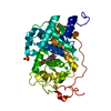 4fcnC  4fcsC  4fdqC  4fefC  2vkaS C: citing same article ( S: Starting model for refinement |
|---|---|
| Similar structure data |


-Links
 PDBj
PDBj
- Assembly
| Deposited unit | 
| ||||||||
|---|---|---|---|---|---|---|---|---|---|
| 1 |
| ||||||||
| Unit cell |
|
-Components
-Protein , 1 types, 1 molecules A
| #1: Protein | Mass: 33318.293 Da / Num. of mol.: 1 / Fragment: RESIDUES 31-347 / Mutation: E140G, G191E Source method: isolated from a genetically manipulated source Source: (gene. exp.) Pleurotus eryngii (fungus) / Gene: vpl2 / Production host:  |
|---|
-Non-polymers , 5 types, 172 molecules 


| #2: Chemical | ChemComp-HEM /  Mass: 616.487 Da / Num. of mol.: 1 / Source method: obtained synthetically / Formula: C34H32FeN4O4 Mass: 616.487 Da / Num. of mol.: 1 / Source method: obtained synthetically / Formula: C34H32FeN4O4 | ||||||
|---|---|---|---|---|---|---|---|
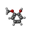
| #3: Chemical |  Mass: 40.078 Da / Num. of mol.: 2 / Source method: obtained synthetically / Formula: Ca Mass: 40.078 Da / Num. of mol.: 2 / Source method: obtained synthetically / Formula: Ca#4: Chemical | ChemComp-JZ3 / |  Mass: 124.137 Da / Num. of mol.: 1 / Source method: obtained synthetically / Formula: C7H8O2 Mass: 124.137 Da / Num. of mol.: 1 / Source method: obtained synthetically / Formula: C7H8O2#5: Chemical | ChemComp-SO4 / |  Mass: 96.063 Da / Num. of mol.: 1 / Source method: obtained synthetically / Formula: SO4 Mass: 96.063 Da / Num. of mol.: 1 / Source method: obtained synthetically / Formula: SO4#6: Water | ChemComp-HOH / | Mass: 18.015 Da / Num. of mol.: 167 / Source method: isolated from a natural source / Formula: H2O |
-Details
| Has protein modification | Y |
|---|
-Experimental details
-Experiment
| Experiment | Method: X-RAY DIFFRACTION / Number of used crystals: 1 |
|---|
- Sample preparation
| Crystal | Density Matthews: 3.45 Å3/Da / Density % sol: 64.31 % |
|---|---|
| Crystal grow | Temperature: 295 K / Method: vapor diffusion, sitting drop / pH: 5 Details: 1.4 M AMMONIUM SULFATE, 0.1 M SODIUM CACODYLATE, PH 5.0, VAPOR DIFFUSION, SITTING DROP, temperature 295K |
-Data collection
| Diffraction | Mean temperature: 100 K |
|---|---|
| Diffraction source | Source: SYNCHROTRON / Site: ESRF  / Beamline: ID14-2 / Wavelength: 0.933 Å / Beamline: ID14-2 / Wavelength: 0.933 Å |
| Detector | Type: ADSC QUANTUM 4r / Detector: CCD / Date: Apr 21, 2008 |
| Radiation | Monochromator: DIAMOND (111) / Protocol: SINGLE WAVELENGTH / Monochromatic (M) / Laue (L): M / Scattering type: x-ray |
| Radiation wavelength | Wavelength: 0.933 Å / Relative weight: 1 |
| Reflection | Resolution: 2.35→69 Å / Num. all: 18866 / Num. obs: 18856 / % possible obs: 100 % / Observed criterion σ(F): 1 / Observed criterion σ(I): 1 / Redundancy: 4.1 % / Biso Wilson estimate: 15.55 Å2 / Rmerge(I) obs: 0.18 / Net I/σ(I): 3.9 |
| Reflection shell | Resolution: 2.35→2.48 Å / Redundancy: 4.1 % / Rmerge(I) obs: 0.454 / Mean I/σ(I) obs: 1.6 / Num. unique all: 2762 / % possible all: 100 |
- Processing
| Software |
| |||||||||||||||||||||||||||||||||||||||||||||
|---|---|---|---|---|---|---|---|---|---|---|---|---|---|---|---|---|---|---|---|---|---|---|---|---|---|---|---|---|---|---|---|---|---|---|---|---|---|---|---|---|---|---|---|---|---|---|
| Refinement | Method to determine structure: MOLECULAR REPLACEMENT Starting model: 2VKA Resolution: 2.35→69 Å / Cor.coef. Fo:Fc: 0.927 / Cor.coef. Fo:Fc free: 0.885 / SU B: 4.934 / SU ML: 0.119 / Cross valid method: THROUGHOUT / ESU R: 0.238 / ESU R Free: 0.198 / Stereochemistry target values: MAXIMUM LIKELIHOOD / Details: HYDROGENS HAVE BEEN USED IF PRESENT IN THE INPUT
| |||||||||||||||||||||||||||||||||||||||||||||
| Solvent computation | Ion probe radii: 0.8 Å / Shrinkage radii: 0.8 Å / VDW probe radii: 1.2 Å / Solvent model: MASK | |||||||||||||||||||||||||||||||||||||||||||||
| Displacement parameters | Biso mean: 8.459 Å2
| |||||||||||||||||||||||||||||||||||||||||||||
| Refinement step | Cycle: LAST / Resolution: 2.35→69 Å
| |||||||||||||||||||||||||||||||||||||||||||||
| Refine LS restraints |
| |||||||||||||||||||||||||||||||||||||||||||||
| LS refinement shell | Resolution: 2.35→2.405 Å / Total num. of bins used: 20
|
