 Movie
Movie Controller

[English] 日本語
 Yorodumi
- PDB-3fm4: Crystal Structure Analysis of Fungal Versatile Peroxidase from Pl... -
+ Open data

- Basic information
| Entry | Database: PDB / ID: 3fm4 | ||||||
|---|---|---|---|---|---|---|---|
| Title | Crystal Structure Analysis of Fungal Versatile Peroxidase from Pleurotus eryngii | ||||||
 Components Components | Versatile peroxidase VPL2 | ||||||
 Keywords Keywords | OXIDOREDUCTASE / CLASS II (FUNGAL) PEROXIDASES / protoporphyrin IX / ELECTRON TRANSFER / LIGNIN PEROXIDASE / LIGNIN DEGRADATION / MANGANESE PEROXIDASE / MN-INDEPENDENT OXIDATION PHENOLIC NON-PHENOLIC AROMATICS / MNII OXIDATION / PEROXIDASE / POLYVALENT PEROXIDASE / Heme / Hydrogen peroxide / Iron / Manganese / Metal-binding / Secreted / Zymogen | ||||||
| Function / homology |  Function and homology information Function and homology informationversatile peroxidase / versatile peroxidase activity / manganese peroxidase activity / lignin catabolic process / hydrogen peroxide catabolic process / response to reactive oxygen species / cellular response to oxidative stress / heme binding / extracellular region / metal ion binding Similarity search - Function | ||||||
| Biological species |  Pleurotus eryngii (fungus) Pleurotus eryngii (fungus) | ||||||
| Method |  X-RAY DIFFRACTION / SYNCHROTRON / MOLECULAR REPLACEMENT / Resolution: 2.11 Å X-RAY DIFFRACTION / SYNCHROTRON / MOLECULAR REPLACEMENT / Resolution: 2.11 Å | ||||||
 Authors Authors | Piontek, K. / Martinez, A.T. / Choinowski, T. / Plattner, D.A. | ||||||
 Citation Citation | Journal: To be Published Title: Structural and Site-directed Mutagenesis Study of Versatile Peroxidase Oxidizing both Mn(II) and Aromatic Substrates Authors: Piontek, K. / Choinowski, T. / Perez-Boada, M. / Ruiz-Duenas, F.J. / Martinez, M.J. / Plattner, D.A. / Martinez, A.T. | ||||||
| History |
|
- Structure visualization
| Structure viewer | Molecule: MolmilJmol/JSmol |
|---|
- Downloads & links
-Download
| PDBx/mmCIF format | 3fm4.cif.gz | 86.4 KB | Display | PDBx/mmCIF format |
|---|---|---|---|---|
| PDB format | pdb3fm4.ent.gz | 64.2 KB | Display | PDB format |
| PDBx/mmJSON format | 3fm4.json.gz | Tree view | PDBx/mmJSON format | |
| Others |  Other downloads Other downloads |
-Validation report
| Arichive directory | https://data.pdbj.org/pub/pdb/validation_reports/fm/3fm4ftp://data.pdbj.org/pub/pdb/validation_reports/fm/3fm4 | HTTPS FTP |
|---|
-Related structure data
| Related structure data |  3fjwC  3fkgSC  3fm1C  3fm6C  3fmuC C: citing same article ( S: Starting model for refinement |
|---|---|
| Similar structure data |


-Links
 PDBj
PDBj


- Assembly
| Deposited unit | 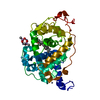
| ||||||||
|---|---|---|---|---|---|---|---|---|---|
| 1 |
| ||||||||
| Unit cell |
|
-Components
-Protein , 1 types, 1 molecules A
| #1: Protein | Mass: 34561.691 Da / Num. of mol.: 1 / Mutation: D175A Source method: isolated from a genetically manipulated source Source: (gene. exp.) Pleurotus eryngii (fungus) / Strain: IJFM, A169 / Gene: VPL2 / Plasmid: PFLAG1-VPL2 / Production host:  |
|---|
-Non-polymers , 6 types, 375 molecules 


| #2: Chemical | ChemComp-HEM /  Mass: 616.487 Da / Num. of mol.: 1 / Source method: obtained synthetically / Formula: C34H32FeN4O4 Mass: 616.487 Da / Num. of mol.: 1 / Source method: obtained synthetically / Formula: C34H32FeN4O4 | ||||||||
|---|---|---|---|---|---|---|---|---|---|
| #3: Chemical |  Mass: 40.078 Da / Num. of mol.: 2 / Source method: obtained synthetically / Formula: Ca Mass: 40.078 Da / Num. of mol.: 2 / Source method: obtained synthetically / Formula: Ca#4: Chemical | ChemComp-ZN /  Mass: 65.409 Da / Num. of mol.: 6 / Source method: obtained synthetically / Formula: Zn Mass: 65.409 Da / Num. of mol.: 6 / Source method: obtained synthetically / Formula: Zn#5: Chemical |  Mass: 55.845 Da / Num. of mol.: 2 / Source method: obtained synthetically / Formula: Fe Mass: 55.845 Da / Num. of mol.: 2 / Source method: obtained synthetically / Formula: Fe#6: Chemical |  Mass: 136.989 Da / Num. of mol.: 2 / Source method: obtained synthetically / Formula: C2H6AsO2 Mass: 136.989 Da / Num. of mol.: 2 / Source method: obtained synthetically / Formula: C2H6AsO2#7: Water | ChemComp-HOH / | Mass: 18.015 Da / Num. of mol.: 362 / Source method: isolated from a natural source / Formula: H2O |
-Details
| Has protein modification | Y |
|---|
-Experimental details
-Experiment
| Experiment | Method: X-RAY DIFFRACTION / Number of used crystals: 1 |
|---|
- Sample preparation
| Crystal | Density Matthews: 2.79 Å3/Da / Density % sol: 55.94 % |
|---|---|
| Crystal grow | Temperature: 298 K / Method: vapor diffusion, hanging drop / pH: 6.5 Details: 9.0mg/ml protein in 10mM Na-tartrate pH 5.5, 16% PEG 8000, 100mM Zn-acetate, 100mM Na-cacodylate pH 6.5, VAPOR DIFFUSION, HANGING DROP, temperature 298K |
-Data collection
| Diffraction | Mean temperature: 100 K |
|---|---|
| Diffraction source | Source: SYNCHROTRON / Site: EMBL/DESY, HAMBURG  / Beamline: X11 / Wavelength: 0.8065 Å / Beamline: X11 / Wavelength: 0.8065 Å |
| Detector | Type: MAR CCD 165 mm / Detector: CCD / Date: Nov 15, 2001 |
| Radiation | Protocol: SINGLE WAVELENGTH / Monochromatic (M) / Laue (L): M / Scattering type: x-ray |
| Radiation wavelength | Wavelength: 0.8065 Å / Relative weight: 1 |
| Reflection | Resolution: 2.11→40 Å / Num. all: 22013 / Num. obs: 21485 / % possible obs: 97.6 % / Observed criterion σ(F): 0 / Observed criterion σ(I): 0 / Redundancy: 2.8 % / Rsym value: 0.084 / Net I/σ(I): 9.1 |
| Reflection shell | Resolution: 2.11→2.15 Å / Mean I/σ(I) obs: 1.7 / Num. unique all: 839 / Rsym value: 0.367 / % possible all: 78.5 |
- Processing
| Software |
| |||||||||||||||||||||||||||||||||||||||||||||||||||||||||||||||||||||||||||||||||||||||||||||||
|---|---|---|---|---|---|---|---|---|---|---|---|---|---|---|---|---|---|---|---|---|---|---|---|---|---|---|---|---|---|---|---|---|---|---|---|---|---|---|---|---|---|---|---|---|---|---|---|---|---|---|---|---|---|---|---|---|---|---|---|---|---|---|---|---|---|---|---|---|---|---|---|---|---|---|---|---|---|---|---|---|---|---|---|---|---|---|---|---|---|---|---|---|---|---|---|---|
| Refinement | Method to determine structure: MOLECULAR REPLACEMENT Starting model: PDB ENTRY 3FKG Resolution: 2.11→32.88 Å / Cor.coef. Fo:Fc: 0.966 / Cor.coef. Fo:Fc free: 0.943 / SU B: 2.415 / SU ML: 0.066 / Cross valid method: THROUGHOUT / σ(F): 0 / ESU R: 0.168 / ESU R Free: 0.158 / Stereochemistry target values: MAXIMUM LIKELIHOOD
| |||||||||||||||||||||||||||||||||||||||||||||||||||||||||||||||||||||||||||||||||||||||||||||||
| Solvent computation | Ion probe radii: 0.8 Å / Shrinkage radii: 0.8 Å / VDW probe radii: 1.2 Å / Solvent model: BABINET MODEL WITH MASK | |||||||||||||||||||||||||||||||||||||||||||||||||||||||||||||||||||||||||||||||||||||||||||||||
| Displacement parameters | Biso mean: 28.046 Å2
| |||||||||||||||||||||||||||||||||||||||||||||||||||||||||||||||||||||||||||||||||||||||||||||||
| Refinement step | Cycle: LAST / Resolution: 2.11→32.88 Å
| |||||||||||||||||||||||||||||||||||||||||||||||||||||||||||||||||||||||||||||||||||||||||||||||
| Refine LS restraints |
| |||||||||||||||||||||||||||||||||||||||||||||||||||||||||||||||||||||||||||||||||||||||||||||||
| LS refinement shell | Resolution: 2.11→2.165 Å / Total num. of bins used: 20
|
