 Movie
Movie Controller

[English] 日本語
 Yorodumi
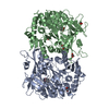
- PDB-4s2a: Crystal structure of Caulobacter crescentus ThiC with Fe4S4 clust... -
+ Open data

- Basic information
| Entry | Database: PDB / ID: 4s2a | ||||||
|---|---|---|---|---|---|---|---|
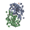
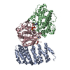
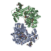
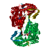
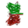
| Title | Crystal structure of Caulobacter crescentus ThiC with Fe4S4 cluster at remote site (holo form) | ||||||
 Components Components | Phosphomethylpyrimidine synthase | ||||||
 Keywords Keywords | LYASE / Alpha-Beta Barrel / Radical SAM superfamily / Iron-sulfur cluster / Thiamin / Vitamin B1 / Vitamin B12 / Domain swapping / AdoMet and Glutamate mutase | ||||||
| Function / homology |  Function and homology information Function and homology informationphosphomethylpyrimidine synthase / phosphomethylpyrimidine synthase activity / thiamine biosynthetic process / thiamine diphosphate biosynthetic process / 4 iron, 4 sulfur cluster binding / zinc ion binding / cytosol Similarity search - Function | ||||||
| Biological species |  Caulobacter crescentus CB15 (bacteria) Caulobacter crescentus CB15 (bacteria) | ||||||
| Method |  X-RAY DIFFRACTION / SYNCHROTRON / MOLECULAR REPLACEMENT / Resolution: 2.93 Å X-RAY DIFFRACTION / SYNCHROTRON / MOLECULAR REPLACEMENT / Resolution: 2.93 Å | ||||||
 Authors Authors | Fenwick, M.K. / Mehta, A.P. / Zhang, Y. / Abdelwahed, S. / Begley, T.P. / Ealick, S.E. | ||||||
 Citation Citation | Journal: Nat Commun Title: Non-canonical active site architecture of the radical SAM thiamin pyrimidine synthase. Authors: Fenwick, M.K. / Mehta, A.P. / Zhang, Y. / Abdelwahed, S.H. / Begley, T.P. / Ealick, S.E. | ||||||
| History |
|
- Structure visualization
| Structure viewer | Molecule: MolmilJmol/JSmol |
|---|
- Downloads & links
-Download
| PDBx/mmCIF format | 4s2a.cif.gz | 125.9 KB | Display | PDBx/mmCIF format |
|---|---|---|---|---|
| PDB format | pdb4s2a.ent.gz | 95.3 KB | Display | PDB format |
| PDBx/mmJSON format | 4s2a.json.gz | Tree view | PDBx/mmJSON format | |
| Others |  Other downloads Other downloads |
-Validation report
| Arichive directory | https://data.pdbj.org/pub/pdb/validation_reports/s2/4s2aftp://data.pdbj.org/pub/pdb/validation_reports/s2/4s2a | HTTPS FTP |
|---|
-Related structure data
| Related structure data |  4s25C  4s26C  4s27C  4s28C  4s29C  3epnS S: Starting model for refinement C: citing same article ( |
|---|---|
| Similar structure data |


-Links
 PDBj
PDBj- Assembly
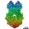
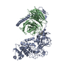
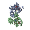
| Deposited unit | 
| ||||||||
|---|---|---|---|---|---|---|---|---|---|
| 1 | 
| ||||||||
| Unit cell |
|
-Components
| #1: Protein | Mass: 68251.336 Da / Num. of mol.: 1 Source method: isolated from a genetically manipulated source Source: (gene. exp.) Caulobacter crescentus CB15 (bacteria) / Strain: ATCC 19089 / CB15 / Gene: thiC, CC_2029 / Plasmid: pET28 / Production host: References: UniProt: Q9A6Q5, phosphomethylpyrimidine synthase |
|---|---|
| #2: Chemical | ChemComp-SF4 /   Mass: 351.640 Da / Num. of mol.: 1 / Source method: obtained synthetically / Formula: Fe4S4 Mass: 351.640 Da / Num. of mol.: 1 / Source method: obtained synthetically / Formula: Fe4S4 |
| #3: Chemical | ChemComp-PO4 /   Mass: 94.971 Da / Num. of mol.: 1 / Source method: obtained synthetically / Formula: PO4 Mass: 94.971 Da / Num. of mol.: 1 / Source method: obtained synthetically / Formula: PO4 |
-Experimental details
-Experiment
| Experiment | Method: X-RAY DIFFRACTION / Number of used crystals: 1 |
|---|
- Sample preparation
| Crystal | Density Matthews: 2.49 Å3/Da / Density % sol: 50.59 % |
|---|---|
| Crystal grow | Temperature: 298 K / Method: vapor diffusion, hanging drop / pH: 9.2 Details: 140 mM HEPES, pH 9.2, and 7.5% (w/v) PEG8000, vapor diffusion, hanging drop, temperature 298K |
-Data collection
| Diffraction | Mean temperature: 100 K | |||||||||||||||||||||||||||||||||||||||||||||||||||||||
|---|---|---|---|---|---|---|---|---|---|---|---|---|---|---|---|---|---|---|---|---|---|---|---|---|---|---|---|---|---|---|---|---|---|---|---|---|---|---|---|---|---|---|---|---|---|---|---|---|---|---|---|---|---|---|---|---|
| Diffraction source | Source: SYNCHROTRON / Site: APS  / Beamline: 24-ID-C / Wavelength: 0.979 Å / Beamline: 24-ID-C / Wavelength: 0.979 Å | |||||||||||||||||||||||||||||||||||||||||||||||||||||||
| Detector | Type: DECTRIS PILATUS 6M-F / Detector: PIXEL / Date: Dec 8, 2012 | |||||||||||||||||||||||||||||||||||||||||||||||||||||||
| Radiation | Protocol: SINGLE WAVELENGTH / Monochromatic (M) / Laue (L): M / Scattering type: x-ray | |||||||||||||||||||||||||||||||||||||||||||||||||||||||
| Radiation wavelength | Wavelength: 0.979 Å / Relative weight: 1 | |||||||||||||||||||||||||||||||||||||||||||||||||||||||
| Reflection | Resolution: 2.93→50 Å / Num. all: 13706 / Num. obs: 13613 / % possible obs: 94.2 % / Observed criterion σ(I): -3 / Redundancy: 2.4 % / Biso Wilson estimate: 33.76 Å2 / Rmerge(I) obs: 0.153 / Net I/σ(I): 5.7 | |||||||||||||||||||||||||||||||||||||||||||||||||||||||
| Reflection shell |
|
- Processing
| Software |
| ||||||||||||||||||||||||||||||||||||||||||
|---|---|---|---|---|---|---|---|---|---|---|---|---|---|---|---|---|---|---|---|---|---|---|---|---|---|---|---|---|---|---|---|---|---|---|---|---|---|---|---|---|---|---|---|
| Refinement | Method to determine structure: MOLECULAR REPLACEMENT Starting model: PDB entry 3EPN Resolution: 2.93→45.55 Å / SU ML: 0.41 / σ(F): 1.37 / Phase error: 25.04 / Stereochemistry target values: ML Details: WEAK RESIDUAL ACTIVE SITE ELECTRON DENSITY IS CONSISTENT WITH LOW OCCUPANCYHMP-P, WHICH WAS NOT ADDED DURING CRYSTALLIZATION BUT WHICH COPURIFIES WITH THE ENZYME. A PHOSPHATE WAS INCLUDED IN ...Details: WEAK RESIDUAL ACTIVE SITE ELECTRON DENSITY IS CONSISTENT WITH LOW OCCUPANCYHMP-P, WHICH WAS NOT ADDED DURING CRYSTALLIZATION BUT WHICH COPURIFIES WITH THE ENZYME. A PHOSPHATE WAS INCLUDED IN THE MODEL, BUT NOT THE PYRIMIDINE
| ||||||||||||||||||||||||||||||||||||||||||
| Solvent computation | Shrinkage radii: 0.9 Å / VDW probe radii: 1.11 Å / Solvent model: FLAT BULK SOLVENT MODEL | ||||||||||||||||||||||||||||||||||||||||||
| Displacement parameters | Biso mean: 26.97 Å2 | ||||||||||||||||||||||||||||||||||||||||||
| Refinement step | Cycle: LAST / Resolution: 2.93→45.55 Å
| ||||||||||||||||||||||||||||||||||||||||||
| Refine LS restraints |
| ||||||||||||||||||||||||||||||||||||||||||
| LS refinement shell |
|
