[English] 日本語
Yorodumi
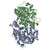
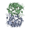
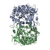
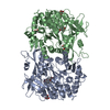
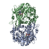
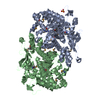
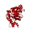
- PDB-4n7q: Crystal structure of eukaryotic THIC from A. thaliana -

+
Open data


ID or keywords:

Loading...

-
Basic information

Entry
Database: PDB / ID: 4n7q
TitleCrystal structure of eukaryotic THIC from A. thaliana
ComponentsPhosphomethylpyrimidine synthase, chloroplastic
KeywordsLYASE / (alpha/beta)8 TIM barrel fold / HMP-P synthase / SAM radical dependent enzyme / metal binding site
Function / homology
Function and homology information


phosphomethylpyrimidine synthase / response to vitamin B1 / carbon-carbon lyase activity / thiamine diphosphate biosynthetic process / thiamine biosynthetic process / plastid / chloroplast stroma / iron-sulfur cluster binding / chloroplast / 4 iron, 4 sulfur cluster binding ...phosphomethylpyrimidine synthase / response to vitamin B1 / carbon-carbon lyase activity / thiamine diphosphate biosynthetic process / thiamine biosynthetic process / plastid / chloroplast stroma / iron-sulfur cluster binding / chloroplast / 4 iron, 4 sulfur cluster binding / protein domain specific binding / metal ion binding / cytosol
Similarity search - Function
Single alpha-helices involved in coiled-coils or other helix-helix interfaces - #620 / Radical SAM ThiC family, central domain / Phosphomethylpyrimidine synthase ThiC/5-hydroxybenzimidazole synthase BzaA/B / Phosphomethylpyrimidine synthase / ThiC/Bza, core domain / Radical SAM ThiC family / Single alpha-helices involved in coiled-coils or other helix-helix interfaces / Helix non-globular / Special / TIM Barrel ...Single alpha-helices involved in coiled-coils or other helix-helix interfaces - #620 / Radical SAM ThiC family, central domain / Phosphomethylpyrimidine synthase ThiC/5-hydroxybenzimidazole synthase BzaA/B / Phosphomethylpyrimidine synthase / ThiC/Bza, core domain / Radical SAM ThiC family / Single alpha-helices involved in coiled-coils or other helix-helix interfaces / Helix non-globular / Special / TIM Barrel / Alpha-Beta Barrel / Alpha Beta
Similarity search - Domain/homology
: / HEXANE-1,6-DIOL / Phosphomethylpyrimidine synthase, chloroplastic
Similarity search - Component
Biological speciesArabidopsis thaliana (thale cress)
MethodX-RAY DIFFRACTION / SYNCHROTRON / MOLECULAR REPLACEMENT / Resolution: 1.6 Å
AuthorsCoquille, S.C. / Roux, C. / Mehta, A. / Begley, T.P. / Fitzpatrick, T.B. / Thore, S.
CitationJournal: J.Struct.Biol. / Year: 2013
Title: High-resolution crystal structure of the eukaryotic HMP-P synthase (THIC) from Arabidopsis thaliana.
Authors: Coquille, S. / Roux, C. / Mehta, A. / Begley, T.P. / Fitzpatrick, T.B. / Thore, S.
History
DepositionOct 16, 2013Deposition site: RCSB / Processing site: RCSB
Revision 1.0Nov 13, 2013Provider: repository / Type: Initial release
Revision 1.1Dec 18, 2013Group: Database references
Revision 1.2Sep 20, 2023Group: Data collection / Database references ...Data collection / Database references / Derived calculations / Refinement description
Category: chem_comp_atom / chem_comp_bond ...chem_comp_atom / chem_comp_bond / database_2 / pdbx_initial_refinement_model / pdbx_struct_conn_angle / struct_conn / struct_ref_seq_dif / struct_site
Item: _database_2.pdbx_DOI / _database_2.pdbx_database_accession ..._database_2.pdbx_DOI / _database_2.pdbx_database_accession / _pdbx_struct_conn_angle.ptnr1_auth_comp_id / _pdbx_struct_conn_angle.ptnr1_auth_seq_id / _pdbx_struct_conn_angle.ptnr1_label_asym_id / _pdbx_struct_conn_angle.ptnr1_label_atom_id / _pdbx_struct_conn_angle.ptnr1_label_comp_id / _pdbx_struct_conn_angle.ptnr1_label_seq_id / _pdbx_struct_conn_angle.ptnr3_auth_comp_id / _pdbx_struct_conn_angle.ptnr3_auth_seq_id / _pdbx_struct_conn_angle.ptnr3_label_asym_id / _pdbx_struct_conn_angle.ptnr3_label_atom_id / _pdbx_struct_conn_angle.ptnr3_label_comp_id / _pdbx_struct_conn_angle.ptnr3_label_seq_id / _pdbx_struct_conn_angle.value / _struct_conn.pdbx_dist_value / _struct_conn.ptnr1_auth_comp_id / _struct_conn.ptnr1_auth_seq_id / _struct_conn.ptnr1_label_asym_id / _struct_conn.ptnr1_label_atom_id / _struct_conn.ptnr1_label_comp_id / _struct_conn.ptnr1_label_seq_id / _struct_conn.ptnr2_auth_comp_id / _struct_conn.ptnr2_auth_seq_id / _struct_conn.ptnr2_label_asym_id / _struct_conn.ptnr2_label_atom_id / _struct_conn.ptnr2_label_comp_id / _struct_ref_seq_dif.details / _struct_site.pdbx_auth_asym_id / _struct_site.pdbx_auth_comp_id / _struct_site.pdbx_auth_seq_id

-
Structure visualization

Structure viewerMolecule:
MolmilJmol/JSmol

Downloads & links

-
Assembly

Deposited unit
A: Phosphomethylpyrimidine synthase, chloroplastic
hetero molecules


Theoretical massNumber of molelcules
Total (without water)65,9605
Polymers65,5461
Non-polymers4134
Water12,520695
1
A: Phosphomethylpyrimidine synthase, chloroplastic
hetero molecules

A: Phosphomethylpyrimidine synthase, chloroplastic
hetero molecules


Theoretical massNumber of molelcules
Total (without water)131,91910
Polymers131,0922
Non-polymers8278
Water362
TypeNameSymmetry operationNumber
identity operation1_555x,y,z1
crystal symmetry operation5_554x-y,-y,-z-2/31
Buried area7080 Å2
ΔGint-50 kcal/mol
Surface area32340 Å2
MethodPISA
Unit cell
Length a, b, c (Å)107.000, 107.000, 88.300
Angle α, β, γ (deg.)90.00, 90.00, 120.00
Int Tables number154
Space group name H-MP3221
Components on special symmetry positions
IDModelComponents
11A-919-

HOH

-
Components

#1: Protein Phosphomethylpyrimidine synthase, chloroplastic / Hydroxymethylpyrimidine phosphate synthase / HMP-P synthase / HMP-phosphate synthase / HMPP ...Hydroxymethylpyrimidine phosphate synthase / HMP-P synthase / HMP-phosphate synthase / HMPP synthase / Protein PYRIMIDINE REQUIRING / Thiamine biosynthesis protein ThiC / Protein THIAMINE C


Mass: 65546.062 Da / Num. of mol.: 1 / Fragment: deltaN71-AtTHIC (unp residues 72-644)
Source method: isolated from a genetically manipulated source
Source: (gene. exp.) Arabidopsis thaliana (thale cress) / Gene: THIC, PY, At2g29630, T27A16.27 / Production host: Escherichia coli (E. coli)
References: UniProt: O82392, phosphomethylpyrimidine synthase
#2: Chemical ChemComp-HEZ / HEXANE-1,6-DIOL


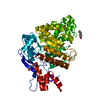
Mass: 118.174 Da / Num. of mol.: 3 / Source method: obtained synthetically / Formula: C6H14O2
#3: Chemical ChemComp-CO / COBALT (II) ION


Mass: 58.933 Da / Num. of mol.: 1 / Source method: obtained synthetically / Formula: Co
#4: Water ChemComp-HOH / water


Mass: 18.015 Da / Num. of mol.: 695 / Source method: isolated from a natural source / Formula: H2O

-
Experimental details

-
Experiment

ExperimentMethod: X-RAY DIFFRACTION / Number of used crystals: 1

-
Sample preparation

CrystalDensity Matthews: 2.23 Å3/Da / Density % sol: 44.75 %
Crystal growTemperature: 291.15 K / Method: vapor diffusion, sitting drop / pH: 4.6
Details: 0.01 M cobalt (II) chloride hexahydrate, 0.1 M sodium acetate trihydrate, 1 M 1,6-Hexanediol, pH 4.6, VAPOR DIFFUSION, SITTING DROP, temperature 291.15K

-
Data collection

DiffractionMean temperature: 100 K
Diffraction sourceSource: SYNCHROTRON / Site: SLS / Beamline: X06DA / Wavelength: 0.97795 Å
DetectorType: DECTRIS PILATUS 2M-F / Detector: PIXEL / Date: Jan 26, 2012
RadiationMonochromator: Bartels Monochromator with dual channel cut crystals (DCCM)
Protocol: SINGLE WAVELENGTH / Monochromatic (M) / Laue (L): M / Scattering type: x-ray
Radiation wavelengthWavelength: 0.97795 Å / Relative weight: 1
ReflectionResolution: 1.6→41.05 Å / Num. all: 77116 / Num. obs: 77101 / % possible obs: 100 % / Redundancy: 10 % / Rmerge(I) obs: 0.146 / Net I/σ(I): 13.58
Reflection shellResolution: 1.6→1.64 Å / Redundancy: 9.95 % / Rmerge(I) obs: 1.271 / Mean I/σ(I) obs: 2.35 / Num. unique all: 5668 / % possible all: 100

-
Processing

Software
NameVersionClassification
XDSdata scaling
CCP4model building
PHENIX(phenix.refine: 1.8.1_1168)refinement
XDSdata reduction
CCP4phasing
RefinementMethod to determine structure: MOLECULAR REPLACEMENT
Starting model: pdb entry 3EPM
Resolution: 1.6→41.027 Å / SU ML: 0.14 / σ(F): 1.99 / Phase error: 17.18 / Stereochemistry target values: ML
RfactorNum. reflection% reflectionSelection details
Rfree0.176 6002 7.79 %RANDOM
Rwork0.1519 ---
obs0.1538 77088 99.97 %-
Solvent computationShrinkage radii: 0.9 Å / VDW probe radii: 1.11 Å / Solvent model: FLAT BULK SOLVENT MODEL
Refinement stepCycle: LAST / Resolution: 1.6→41.027 Å
ProteinNucleic acidLigandSolventTotal
Num. atoms3752 0 25 695 4472
Refine LS restraints
Refine-IDTypeDev idealNumber
X-RAY DIFFRACTIONf_bond_d0.0073945
X-RAY DIFFRACTIONf_angle_d1.0685355
X-RAY DIFFRACTIONf_dihedral_angle_d13.5471464
X-RAY DIFFRACTIONf_chiral_restr0.078570
X-RAY DIFFRACTIONf_plane_restr0.005701
LS refinement shell
Resolution (Å)Rfactor RfreeNum. reflection RfreeRfactor RworkNum. reflection RworkRefine-ID% reflection obs (%)
1.6-1.61820.25781980.24362332X-RAY DIFFRACTION100
1.6182-1.63720.26472000.23372344X-RAY DIFFRACTION100
1.6372-1.65720.28251980.23182335X-RAY DIFFRACTION100
1.6572-1.67820.24111980.22432356X-RAY DIFFRACTION100
1.6782-1.70020.25522010.22082355X-RAY DIFFRACTION100
1.7002-1.72350.26061960.20852331X-RAY DIFFRACTION100
1.7235-1.74820.23732010.19872361X-RAY DIFFRACTION100
1.7482-1.77430.20831960.19882342X-RAY DIFFRACTION100
1.7743-1.8020.21682000.19332370X-RAY DIFFRACTION100
1.802-1.83150.24811960.19582330X-RAY DIFFRACTION100
1.8315-1.86310.21990.1882353X-RAY DIFFRACTION100
1.8631-1.8970.22091980.18192356X-RAY DIFFRACTION100
1.897-1.93350.20832000.18062354X-RAY DIFFRACTION100
1.9335-1.97290.22231990.16812361X-RAY DIFFRACTION100
1.9729-2.01580.1882000.16422373X-RAY DIFFRACTION100
2.0158-2.06270.17651970.16122366X-RAY DIFFRACTION100
2.0627-2.11430.17851990.15442338X-RAY DIFFRACTION100
2.1143-2.17150.15331970.15252355X-RAY DIFFRACTION100
2.1715-2.23540.18641990.15112376X-RAY DIFFRACTION100
2.2354-2.30750.19731990.15582372X-RAY DIFFRACTION100
2.3075-2.390.16461970.15072371X-RAY DIFFRACTION100
2.39-2.48570.18321960.14332384X-RAY DIFFRACTION100
2.4857-2.59880.15692010.13842370X-RAY DIFFRACTION100
2.5988-2.73570.1632010.13782364X-RAY DIFFRACTION100
2.7357-2.90710.17081980.14022400X-RAY DIFFRACTION100
2.9071-3.13150.16492010.1362387X-RAY DIFFRACTION100
3.1315-3.44650.16162040.1352398X-RAY DIFFRACTION100
3.4465-3.94480.1312090.1222404X-RAY DIFFRACTION100
3.9448-4.96870.12382040.10792430X-RAY DIFFRACTION100
4.9687-41.04090.14572200.1282518X-RAY DIFFRACTION100

+
About Yorodumi

-
News

-
Feb 9, 2022. New format data for meta-information of EMDB entries

New format data for meta-information of EMDB entries

  • Version 3 of the EMDB header file is now the official format.
  • The previous official version 1.9 will be removed from the archive.

Related info.:EMDB header

External links:wwPDB to switch to version 3 of the EMDB data model

-
Aug 12, 2020. Covid-19 info

Covid-19 info

URL: https://pdbjlvh1.pdbj.org/emnavi/covid19.php

New page: Covid-19 featured information page in EM Navigator.

Related info.:Covid-19 info / Mar 5, 2020. Novel coronavirus structure data

+
Mar 5, 2020. Novel coronavirus structure data

Novel coronavirus structure data

Related info.:Yorodumi Speices / Aug 12, 2020. Covid-19 info

External links:COVID-19 featured content - PDBj / Molecule of the Month (242):Coronavirus Proteases

+
Jan 31, 2019. EMDB accession codes are about to change! (news from PDBe EMDB page)

EMDB accession codes are about to change! (news from PDBe EMDB page)

  • The allocation of 4 digits for EMDB accession codes will soon come to an end. Whilst these codes will remain in use, new EMDB accession codes will include an additional digit and will expand incrementally as the available range of codes is exhausted. The current 4-digit format prefixed with “EMD-” (i.e. EMD-XXXX) will advance to a 5-digit format (i.e. EMD-XXXXX), and so on. It is currently estimated that the 4-digit codes will be depleted around Spring 2019, at which point the 5-digit format will come into force.
  • The EM Navigator/Yorodumi systems omit the EMD- prefix.

Related info.:Q: What is EMD? / ID/Accession-code notation in Yorodumi/EM Navigator

External links:EMDB Accession Codes are Changing Soon! / Contact to PDBj

+
Jul 12, 2017. Major update of PDB

Major update of PDB

  • wwPDB released updated PDB data conforming to the new PDBx/mmCIF dictionary.
  • This is a major update changing the version number from 4 to 5, and with Remediation, in which all the entries are updated.
  • In this update, many items about electron microscopy experimental information are reorganized (e.g. em_software).
  • Now, EM Navigator and Yorodumi are based on the updated data.

External links:wwPDB Remediation / Enriched Model Files Conforming to OneDep Data Standards Now Available in the PDB FTP Archive

-
Yorodumi

Thousand views of thousand structures

  • Yorodumi is a browser for structure data from EMDB, PDB, SASBDB, etc.
  • This page is also the successor to EM Navigator detail page, and also detail information page/front-end page for Omokage search.
  • The word "yorodu" (or yorozu) is an old Japanese word meaning "ten thousand". "mi" (miru) is to see.

Related info.:EMDB / PDB / SASBDB / Comparison of 3 databanks / Yorodumi Search / Aug 31, 2016. New EM Navigator & Yorodumi / Yorodumi Papers / Jmol/JSmol / Function and homology information / Changes in new EM Navigator and Yorodumi

Read more