[English] 日本語
Yorodumi
- PDB-4lac: Crystal Structure of Protein Phosphatase 2A (PP2A) and PP2A phosp... -

+
Open data


ID or keywords:

Loading...

-
Basic information

Entry
Database: PDB / ID: 4lac
TitleCrystal Structure of Protein Phosphatase 2A (PP2A) and PP2A phosphatase activator (PTPA) complex with ATPgammaS
Components
  • (Serine/threonine-protein phosphatase 2A ...) x 2
  • PP2A Scaffold Subunit A, Truncated, an internal deletion of PP2A A
KeywordsHydrolase/signaling protein / PP2A / PTPA / protein phosphatase / signaling pathway regulation / chaperone / Hydrolase-signaling protein complex
Function / homology
Function and homology information


protein tyrosine phosphatase activator activity / negative regulation of protein dephosphorylation / meiotic spindle elongation / Integration of energy metabolism / regulation of phosphoprotein phosphatase activity / PP2A-mediated dephosphorylation of key metabolic factors / regulation of microtubule binding / MASTL Facilitates Mitotic Progression / mitotic sister chromatid separation / regulation of meiotic cell cycle process involved in oocyte maturation ...protein tyrosine phosphatase activator activity / negative regulation of protein dephosphorylation / meiotic spindle elongation / Integration of energy metabolism / regulation of phosphoprotein phosphatase activity / PP2A-mediated dephosphorylation of key metabolic factors / regulation of microtubule binding / MASTL Facilitates Mitotic Progression / mitotic sister chromatid separation / regulation of meiotic cell cycle process involved in oocyte maturation / protein phosphatase type 2A complex / meiotic sister chromatid cohesion, centromeric / peptidyl-serine dephosphorylation / peptidyl-threonine dephosphorylation / : / positive regulation of microtubule binding / negative regulation of tyrosine phosphorylation of STAT protein / Regulation of glycolysis by fructose 2,6-bisphosphate metabolism / Inhibition of replication initiation of damaged DNA by RB1/E2F1 / female meiotic nuclear division / protein antigen binding / protein phosphatase regulator activity / ceramide metabolic process / GABA receptor binding / negative regulation of epithelial to mesenchymal transition / APC truncation mutants have impaired AXIN binding / AXIN missense mutants destabilize the destruction complex / Truncations of AMER1 destabilize the destruction complex / Initiation of Nuclear Envelope (NE) Reformation / ERKs are inactivated / positive regulation of extrinsic apoptotic signaling pathway in absence of ligand / Beta-catenin phosphorylation cascade / Signaling by GSK3beta mutants / CTNNB1 S33 mutants aren't phosphorylated / CTNNB1 S37 mutants aren't phosphorylated / CTNNB1 S45 mutants aren't phosphorylated / CTNNB1 T41 mutants aren't phosphorylated / regulation of Wnt signaling pathway / Disassembly of the destruction complex and recruitment of AXIN to the membrane / regulation of growth / negative regulation of glycolytic process through fructose-6-phosphate / positive regulation of NLRP3 inflammasome complex assembly / myosin phosphatase activity / protein serine/threonine phosphatase activity / ATPase complex / negative regulation of phosphoprotein phosphatase activity / CTLA4 inhibitory signaling / Platelet sensitization by LDL / negative regulation of MAPK cascade / protein-serine/threonine phosphatase / regulation of cell differentiation / T cell homeostasis / ERK/MAPK targets / regulation of G1/S transition of mitotic cell cycle / regulation of DNA replication / phosphoprotein phosphatase activity / mesoderm development / positive regulation of phosphoprotein phosphatase activity / chromosome, centromeric region / DARPP-32 events / negative regulation of phosphatidylinositol 3-kinase/protein kinase B signal transduction / lateral plasma membrane / Nonsense Mediated Decay (NMD) enhanced by the Exon Junction Complex (EJC) / Amplification of signal from unattached kinetochores via a MAD2 inhibitory signal / regulation of cell adhesion / Cyclin A/B1/B2 associated events during G2/M transition / Mitotic Prometaphase / EML4 and NUDC in mitotic spindle formation / positive regulation of protein dephosphorylation / Loss of Nlp from mitotic centrosomes / Loss of proteins required for interphase microtubule organization from the centrosome / Recruitment of mitotic centrosome proteins and complexes / Resolution of Sister Chromatid Cohesion / Recruitment of NuMA to mitotic centrosomes / calcium channel complex / Anchoring of the basal body to the plasma membrane / AURKA Activation by TPX2 / protein dephosphorylation / mitotic spindle organization / RNA splicing / meiotic cell cycle / protein phosphatase 2A binding / peptidylprolyl isomerase / response to organic substance / protein tyrosine phosphatase activity / peptidyl-prolyl cis-trans isomerase activity / chromosome segregation / RHO GTPases Activate Formins / response to lead ion / regulation of protein phosphorylation / Spry regulation of FGF signaling / RAF activation / tau protein binding / PKR-mediated signaling / Degradation of beta-catenin by the destruction complex / positive regulation of protein serine/threonine kinase activity / negative regulation of cell growth / spindle pole / Negative regulation of MAPK pathway / Separation of Sister Chromatids
Similarity search - Function
Phosphotyrosyl phosphate activator, C-terminal lid domain / Phosphotyrosyl phosphatase activator, PTPA / PTPA superfamily / Phosphotyrosyl phosphatase activator, C-terminal lid domain / Phosphotyrosyl phosphate activator (PTPA) protein / : / HEAT repeat / HEAT repeat / Serine/threonine specific protein phosphatases signature. / Protein phosphatase 2A homologues, catalytic domain. ...Phosphotyrosyl phosphate activator, C-terminal lid domain / Phosphotyrosyl phosphatase activator, PTPA / PTPA superfamily / Phosphotyrosyl phosphatase activator, C-terminal lid domain / Phosphotyrosyl phosphate activator (PTPA) protein / : / HEAT repeat / HEAT repeat / Serine/threonine specific protein phosphatases signature. / Protein phosphatase 2A homologues, catalytic domain. / Serine/threonine-specific protein phosphatase/bis(5-nucleosyl)-tetraphosphatase / HEAT repeat profile. / HEAT, type 2 / HEAT repeats / Metallo-dependent phosphatases / Purple Acid Phosphatase; chain A, domain 2 / Calcineurin-like phosphoesterase domain, ApaH type / Calcineurin-like phosphoesterase / Leucine-rich Repeat Variant / Leucine-rich Repeat Variant / Metallo-dependent phosphatase-like / Four Helix Bundle (Hemerythrin (Met), subunit A) / Armadillo-like helical / 4-Layer Sandwich / Alpha Horseshoe / Armadillo-type fold / Up-down Bundle / Mainly Alpha / Alpha Beta
Similarity search - Domain/homology
PHOSPHOTHIOPHOSPHORIC ACID-ADENYLATE ESTER / : / DI(HYDROXYETHYL)ETHER / Serine/threonine-protein phosphatase 2A 65 kDa regulatory subunit A alpha isoform / Serine/threonine-protein phosphatase 2A catalytic subunit alpha isoform / Serine/threonine-protein phosphatase 2A activator
Similarity search - Component
Biological speciesHomo sapiens (human)
MethodX-RAY DIFFRACTION / SYNCHROTRON / MOLECULAR REPLACEMENT / Resolution: 2.82 Å
AuthorsGuo, F. / Stanevich, V. / Wlodarchak, N. / Satyshur, K.A. / Xing, Y.
CitationJournal: Cell Res. / Year: 2014
Title: Structural basis of PP2A activation by PTPA, an ATP-dependent activation chaperone.
Authors: Guo, F. / Stanevich, V. / Wlodarchak, N. / Sengupta, R. / Jiang, L. / Satyshur, K.A. / Xing, Y.
History
DepositionJun 19, 2013Deposition site: RCSB / Processing site: RCSB
Revision 1.0Oct 9, 2013Provider: repository / Type: Initial release
Revision 1.1Oct 30, 2013Group: Database references
Revision 1.2Feb 19, 2014Group: Database references
Revision 1.3Sep 20, 2023Group: Data collection / Database references ...Data collection / Database references / Derived calculations / Refinement description
Category: chem_comp_atom / chem_comp_bond ...chem_comp_atom / chem_comp_bond / database_2 / pdbx_initial_refinement_model / pdbx_struct_conn_angle / struct_conn / struct_ref_seq_dif / struct_site
Item: _database_2.pdbx_DOI / _database_2.pdbx_database_accession ..._database_2.pdbx_DOI / _database_2.pdbx_database_accession / _pdbx_struct_conn_angle.ptnr1_auth_comp_id / _pdbx_struct_conn_angle.ptnr1_auth_seq_id / _pdbx_struct_conn_angle.ptnr1_label_atom_id / _pdbx_struct_conn_angle.ptnr1_label_comp_id / _pdbx_struct_conn_angle.ptnr1_label_seq_id / _pdbx_struct_conn_angle.ptnr3_auth_comp_id / _pdbx_struct_conn_angle.ptnr3_auth_seq_id / _pdbx_struct_conn_angle.ptnr3_label_atom_id / _pdbx_struct_conn_angle.ptnr3_label_comp_id / _pdbx_struct_conn_angle.ptnr3_label_seq_id / _pdbx_struct_conn_angle.value / _struct_conn.pdbx_dist_value / _struct_conn.ptnr1_auth_comp_id / _struct_conn.ptnr1_auth_seq_id / _struct_conn.ptnr1_label_asym_id / _struct_conn.ptnr1_label_atom_id / _struct_conn.ptnr1_label_comp_id / _struct_conn.ptnr1_label_seq_id / _struct_conn.ptnr2_auth_comp_id / _struct_conn.ptnr2_auth_seq_id / _struct_conn.ptnr2_label_asym_id / _struct_conn.ptnr2_label_atom_id / _struct_conn.ptnr2_label_comp_id / _struct_ref_seq_dif.details / _struct_site.pdbx_auth_asym_id / _struct_site.pdbx_auth_comp_id / _struct_site.pdbx_auth_seq_id

-
Structure visualization

Structure viewerMolecule:
MolmilJmol/JSmol

Downloads & links

-
Assembly

Deposited unit
B: Serine/threonine-protein phosphatase 2A activator
A: PP2A Scaffold Subunit A, Truncated, an internal deletion of PP2A A
C: Serine/threonine-protein phosphatase 2A catalytic subunit alpha isoform
hetero molecules


Theoretical massNumber of molelcules
Total (without water)100,3998
Polymers99,4643
Non-polymers9345
Water3,153175
1


  • Idetical with deposited unit
  • defined by author
TypeNameSymmetry operationNumber
identity operation1_555x,y,z1
Unit cell
Length a, b, c (Å)57.070, 100.186, 167.450
Angle α, β, γ (deg.)90.00, 90.00, 90.00
Int Tables number19
Space group name H-MP212121

-
Components

-
Serine/threonine-protein phosphatase 2A ... , 2 types, 2 molecules BC

#1: Protein Serine/threonine-protein phosphatase 2A activator / PP2A / subunit B' / PR53 isoform / Phosphotyrosyl phosphatase activator / PTPA / Serine/threonine- ...PP2A / subunit B' / PR53 isoform / Phosphotyrosyl phosphatase activator / PTPA / Serine/threonine-protein phosphatase 2A regulatory subunit 4 / Serine/threonine-protein phosphatase 2A regulatory subunit B'


Mass: 35187.457 Da / Num. of mol.: 1 / Fragment: unp residues 19-358
Source method: isolated from a genetically manipulated source
Source: (gene. exp.) Homo sapiens (human) / Gene: PP2A Catalytic Subunit C, PPP2R4, PTPA / Plasmid: FBQH / Cell line (production host): Hi5 / Production host: Trichoplusia ni (cabbage looper) / References: UniProt: Q15257, peptidylprolyl isomerase
#3: Protein Serine/threonine-protein phosphatase 2A catalytic subunit alpha isoform / PP2A-alpha / Replication protein C / RP-C


Mass: 35780.281 Da / Num. of mol.: 1
Source method: isolated from a genetically manipulated source
Source: (gene. exp.) Homo sapiens (human) / Gene: PP2A Scaffold Subunit A, PPP2CA / Plasmid: pQlink / Production host: Escherichia coli (E. coli) / Strain (production host): DH5alpha
References: UniProt: P67775, protein-serine/threonine phosphatase

-
Protein , 1 types, 1 molecules A

#2: Protein PP2A Scaffold Subunit A, Truncated, an internal deletion of PP2A A


Mass: 28496.668 Da / Num. of mol.: 1
Source method: isolated from a genetically manipulated source
Source: (gene. exp.) Homo sapiens (human) / Gene: PP2A Phosphatase Activator / Plasmid: pQlink / Production host: Escherichia coli (E. coli) / Strain (production host): DH5alpha / References: UniProt: P30153*PLUS

-
Non-polymers , 5 types, 180 molecules

#4: Chemical ChemComp-PEG / DI(HYDROXYETHYL)ETHER / Diethylene glycol


Mass: 106.120 Da / Num. of mol.: 1 / Source method: obtained synthetically / Formula: C4H10O3
#5: Chemical ChemComp-MN / MANGANESE (II) ION


Mass: 54.938 Da / Num. of mol.: 2 / Source method: obtained synthetically / Formula: Mn
#6: Chemical ChemComp-AGS / PHOSPHOTHIOPHOSPHORIC ACID-ADENYLATE ESTER / ATP-GAMMA-S / ADENOSINE 5'-(3-THIOTRIPHOSPHATE) / ADENOSINE 5'-(GAMMA-THIOTRIPHOSPHATE) / ADENOSINE-5'-DIPHOSPHATE MONOTHIOPHOSPHATE


Mass: 523.247 Da / Num. of mol.: 1 / Source method: obtained synthetically / Formula: C10H16N5O12P3S / Comment: ATP-gamma-S, energy-carrying molecule analogue*YM
#7: Chemical ChemComp-MES / 2-(N-MORPHOLINO)-ETHANESULFONIC ACID / MES (buffer)


Mass: 195.237 Da / Num. of mol.: 1 / Source method: obtained synthetically / Formula: C6H13NO4S / Comment: pH buffer*YM
#8: Water ChemComp-HOH / water / Water


Mass: 18.015 Da / Num. of mol.: 175 / Source method: isolated from a natural source / Formula: H2O

-
Details

Sequence details
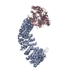
CHAIN A IS PP2A A SUBUNIT WITH INTERNAL DELETIONS OF RESIDUE 2-8 AND 55-407, AND WITH A MUTATION OF F438Y

-
Experimental details

-
Experiment

ExperimentMethod: X-RAY DIFFRACTION / Number of used crystals: 1

-
Sample preparation

CrystalDensity Matthews: 2.41 Å3/Da / Density % sol: 48.89 %
Crystal growTemperature: 298 K / Method: vapor diffusion, sitting drop / pH: 6.5
Details: 0.1M MES at pH6.5 and 10-12% PEG20,000, 1mM ATPgammaS, VAPOR DIFFUSION, SITTING DROP, temperature 298K

-
Data collection

DiffractionMean temperature: 100 K
Diffraction sourceSource: SYNCHROTRON / Site: APS / Beamline: 21-ID-G / Wavelength: 0.97856 Å
DetectorType: MARMOSAIC 300 mm CCD / Detector: CCD / Date: Oct 24, 2009
RadiationProtocol: SINGLE WAVELENGTH / Monochromatic (M) / Laue (L): M / Scattering type: x-ray
Radiation wavelengthWavelength: 0.97856 Å / Relative weight: 1
ReflectionResolution: 2.8→50 Å / Num. all: 23884 / Num. obs: 23656 / % possible obs: 99.1 % / Observed criterion σ(F): 0 / Observed criterion σ(I): 0 / Redundancy: 7.5 % / Rsym value: 0.15 / Net I/σ(I): 11.1
Reflection shellResolution: 2.82→2.87 Å / Redundancy: 4.5 % / Mean I/σ(I) obs: 2.02 / Num. unique all: 1060 / Rsym value: 0.561 / % possible all: 90.4

-
Processing

Software
NameVersionClassification
ADSCQuantumdata collection
PHASERMRphasing
REFMAC5.6.0119refinement
HKL-2000data reduction
HKL-2000data scaling
RefinementMethod to determine structure: MOLECULAR REPLACEMENT
Starting model: PDB ENTRY 3C5W PDB ENTRY 2IE3 PDB ENTRY 2HV6
Resolution: 2.82→50 Å / Cor.coef. Fo:Fc: 0.932 / Cor.coef. Fo:Fc free: 0.894 / SU B: 29.219 / SU ML: 0.284 / Cross valid method: THROUGHOUT / σ(F): 0 / ESU R Free: 0.385 / Stereochemistry target values: MAXIMUM LIKELIHOOD / Details: HYDROGENS HAVE BEEN USED IF PRESENT IN THE INPUT
RfactorNum. reflection% reflectionSelection details
Rfree0.24226 1209 5.1 %RANDOM
Rwork0.18767 ---
obs0.19057 22394 98.91 %-
all-22394 --
Solvent computationIon probe radii: 0.8 Å / Shrinkage radii: 0.8 Å / VDW probe radii: 1.2 Å / Solvent model: BABINET MODEL WITH MASK
Displacement parametersBiso mean: 37.174 Å2
Baniso -1Baniso -2Baniso -3
1-0.94 Å20 Å20 Å2
2---0.53 Å20 Å2
3----0.4 Å2
Refinement stepCycle: LAST / Resolution: 2.82→50 Å
ProteinNucleic acidLigandSolventTotal
Num. atoms6586 0 52 175 6813
Refine LS restraints
Refine-IDTypeDev idealDev ideal targetNumber
X-RAY DIFFRACTIONr_bond_refined_d0.0060.026791
X-RAY DIFFRACTIONr_angle_refined_deg1.1651.9669217
X-RAY DIFFRACTIONr_dihedral_angle_1_deg4.8245817
X-RAY DIFFRACTIONr_dihedral_angle_2_deg39.24824.19315
X-RAY DIFFRACTIONr_dihedral_angle_3_deg15.719151170
X-RAY DIFFRACTIONr_dihedral_angle_4_deg17.461538
X-RAY DIFFRACTIONr_chiral_restr0.0930.21024
X-RAY DIFFRACTIONr_gen_planes_refined0.0040.0215098
LS refinement shellResolution: 2.822→2.895 Å / Total num. of bins used: 20
RfactorNum. reflection% reflection
Rfree0.332 71 -
Rwork0.282 1339 -
obs--89.24 %
Refinement TLS params.

Method: refined / Refine-ID: X-RAY DIFFRACTION

IDL112)L122)L132)L222)L232)L332)S11 (Å °)S12 (Å °)S13 (Å °)S21 (Å °)S22 (Å °)S23 (Å °)S31 (Å °)S32 (Å °)S33 (Å °)T112)T122)T132)T222)T232)T332)Origin x (Å)Origin y (Å)Origin z (Å)
10.58380.20040.07980.8606-0.16990.6126-0.03880.01970.0313-0.00110.0919-0.0373-0.05190.0647-0.05310.28170.00390.02760.03880.00440.0358-13.03025.440217.6679
22.7993-0.1637-1.67370.62470.14041.00560.0746-0.0332-0.0873-0.1677-0.11650.0271-0.04280.00970.0420.30160.0246-0.0160.04260.00320.0152-29.2267-48.987819.7234
30.60910.09860.23050.89540.1260.70430.0268-0.0044-0.0126-0.02510.00670.0352-0.00710.0133-0.03350.28040.0131-0.00910.0305-0.00160.0469-34.0525-17.860529.8401
Refinement TLS group
IDRefine-IDRefine TLS-IDAuth asym-IDAuth seq-ID
1X-RAY DIFFRACTION1B21 - 321
2X-RAY DIFFRACTION2A360 - 588
3X-RAY DIFFRACTION3C4 - 293
4X-RAY DIFFRACTION3C501 - 502

+
About Yorodumi

-
News

-
Feb 9, 2022. New format data for meta-information of EMDB entries

New format data for meta-information of EMDB entries

  • Version 3 of the EMDB header file is now the official format.
  • The previous official version 1.9 will be removed from the archive.

Related info.:EMDB header

External links:wwPDB to switch to version 3 of the EMDB data model

-
Aug 12, 2020. Covid-19 info

Covid-19 info

URL: https://pdbj.org/emnavi/covid19.php

New page: Covid-19 featured information page in EM Navigator.

Related info.:Covid-19 info / Mar 5, 2020. Novel coronavirus structure data

+
Mar 5, 2020. Novel coronavirus structure data

Novel coronavirus structure data

Related info.:Yorodumi Speices / Aug 12, 2020. Covid-19 info

External links:COVID-19 featured content - PDBj / Molecule of the Month (242):Coronavirus Proteases

+
Jan 31, 2019. EMDB accession codes are about to change! (news from PDBe EMDB page)

EMDB accession codes are about to change! (news from PDBe EMDB page)

  • The allocation of 4 digits for EMDB accession codes will soon come to an end. Whilst these codes will remain in use, new EMDB accession codes will include an additional digit and will expand incrementally as the available range of codes is exhausted. The current 4-digit format prefixed with “EMD-” (i.e. EMD-XXXX) will advance to a 5-digit format (i.e. EMD-XXXXX), and so on. It is currently estimated that the 4-digit codes will be depleted around Spring 2019, at which point the 5-digit format will come into force.
  • The EM Navigator/Yorodumi systems omit the EMD- prefix.

Related info.:Q: What is EMD? / ID/Accession-code notation in Yorodumi/EM Navigator

External links:EMDB Accession Codes are Changing Soon! / Contact to PDBj

+
Jul 12, 2017. Major update of PDB

Major update of PDB

  • wwPDB released updated PDB data conforming to the new PDBx/mmCIF dictionary.
  • This is a major update changing the version number from 4 to 5, and with Remediation, in which all the entries are updated.
  • In this update, many items about electron microscopy experimental information are reorganized (e.g. em_software).
  • Now, EM Navigator and Yorodumi are based on the updated data.

External links:wwPDB Remediation / Enriched Model Files Conforming to OneDep Data Standards Now Available in the PDB FTP Archive

-
Yorodumi

Thousand views of thousand structures

  • Yorodumi is a browser for structure data from EMDB, PDB, SASBDB, etc.
  • This page is also the successor to EM Navigator detail page, and also detail information page/front-end page for Omokage search.
  • The word "yorodu" (or yorozu) is an old Japanese word meaning "ten thousand". "mi" (miru) is to see.

Related info.:EMDB / PDB / SASBDB / Comparison of 3 databanks / Yorodumi Search / Aug 31, 2016. New EM Navigator & Yorodumi / Yorodumi Papers / Jmol/JSmol / Function and homology information / Changes in new EM Navigator and Yorodumi

Read more