 Movie
Movie Controller

[English] 日本語
 Yorodumi
- PDB-3c5w: Complex between PP2A-specific methylesterase PME-1 and PP2A core ... -
+ Open data

- Basic information
| Entry | Database: PDB / ID: 3c5w | ||||||
|---|---|---|---|---|---|---|---|
| Title | Complex between PP2A-specific methylesterase PME-1 and PP2A core enzyme | ||||||
 Components Components |
| ||||||
 Keywords Keywords | HYDROLASE / methylesterase / phosphatase / PP2A | ||||||
| Function / homology |  Function and homology information Function and homology informationprotein phosphatase methylesterase-1 / protein methylesterase activity / meiotic spindle elongation / PP2A-mediated dephosphorylation of key metabolic factors / RNA polymerase II CTD heptapeptide repeat S2 phosphatase activity / RNA polymerase II CTD heptapeptide repeat S7 phosphatase activity / peptidyl-threonine dephosphorylation / regulation of meiotic cell cycle process involved in oocyte maturation / mitotic sister chromatid separation / MASTL Facilitates Mitotic Progression ...protein phosphatase methylesterase-1 / protein methylesterase activity / meiotic spindle elongation / PP2A-mediated dephosphorylation of key metabolic factors / RNA polymerase II CTD heptapeptide repeat S2 phosphatase activity / RNA polymerase II CTD heptapeptide repeat S7 phosphatase activity / peptidyl-threonine dephosphorylation / regulation of meiotic cell cycle process involved in oocyte maturation / mitotic sister chromatid separation / MASTL Facilitates Mitotic Progression / protein phosphatase type 2A complex / meiotic sister chromatid cohesion, centromeric / INTAC complex / RNA polymerase II CTD heptapeptide repeat S5 phosphatase activity / FAR/SIN/STRIPAK complex / female meiotic nuclear division / Regulation of glycolysis by fructose 2,6-bisphosphate metabolism / Inhibition of replication initiation of damaged DNA by RB1/E2F1 / protein phosphatase regulator activity / protein antigen binding / GABA receptor binding / APC truncation mutants have impaired AXIN binding / AXIN missense mutants destabilize the destruction complex / Truncations of AMER1 destabilize the destruction complex / positive regulation of extrinsic apoptotic signaling pathway in absence of ligand / ERKs are inactivated / Initiation of Nuclear Envelope (NE) Reformation / Beta-catenin phosphorylation cascade / Signaling by GSK3beta mutants / CTNNB1 S33 mutants aren't phosphorylated / CTNNB1 S37 mutants aren't phosphorylated / CTNNB1 S45 mutants aren't phosphorylated / CTNNB1 T41 mutants aren't phosphorylated / Co-stimulation by CD28 / regulation of growth / RNA polymerase II transcription initiation surveillance / Disassembly of the destruction complex and recruitment of AXIN to the membrane / protein dephosphorylation / negative regulation of epithelial to mesenchymal transition / Co-inhibition by CTLA4 / protein phosphatase inhibitor activity / Platelet sensitization by LDL / protein-serine/threonine phosphatase / ERK/MAPK targets / negative regulation of glycolytic process through fructose-6-phosphate / lncRNA binding / vascular endothelial cell response to oscillatory fluid shear stress / mesoderm development / protein serine/threonine phosphatase activity / T cell homeostasis / positive regulation of NLRP3 inflammasome complex assembly / regulation of cell differentiation / regulation of microtubule polymerization / regulation of G1/S transition of mitotic cell cycle / lateral plasma membrane / chromosome, centromeric region / DARPP-32 events / negative regulation of hippo signaling / Cyclin A/B1/B2 associated events during G2/M transition / Nonsense Mediated Decay (NMD) enhanced by the Exon Junction Complex (EJC) / spindle assembly / phosphoprotein phosphatase activity / Amplification of signal from unattached kinetochores via a MAD2 inhibitory signal / Loss of Nlp from mitotic centrosomes / Loss of proteins required for interphase microtubule organization from the centrosome / Recruitment of mitotic centrosome proteins and complexes / protein tyrosine phosphatase activity / Recruitment of NuMA to mitotic centrosomes / Anchoring of the basal body to the plasma membrane / Mitotic Prometaphase / EML4 and NUDC in mitotic spindle formation / protein phosphatase 2A binding / Turbulent (oscillatory, disturbed) flow shear stress activates signaling by PIEZO1 and integrins in endothelial cells / AURKA Activation by TPX2 / Resolution of Sister Chromatid Cohesion / negative regulation of phosphatidylinositol 3-kinase/protein kinase B signal transduction / meiotic cell cycle / chromosome segregation / negative regulation of canonical Wnt signaling pathway / RAF activation / RHO GTPases Activate Formins / Spry regulation of FGF signaling / PKR-mediated signaling / response to lead ion / Degradation of beta-catenin by the destruction complex / G2/M transition of mitotic cell cycle / tau protein binding / spindle pole / Cyclin D associated events in G1 / Negative regulation of MAPK pathway / Separation of Sister Chromatids / Regulation of TP53 Degradation / Regulation of PLK1 Activity at G2/M Transition / mitotic cell cycle / PI5P, PP2A and IER3 Regulate PI3K/AKT Signaling / microtubule cytoskeleton / protein-containing complex assembly / protein phosphatase binding / neuron projection / intracellular signal transduction Similarity search - Function | ||||||
| Biological species |  Homo sapiens (human) Homo sapiens (human) | ||||||
| Method |  X-RAY DIFFRACTION / SYNCHROTRON / MOLECULAR REPLACEMENT / Resolution: 2.8 Å X-RAY DIFFRACTION / SYNCHROTRON / MOLECULAR REPLACEMENT / Resolution: 2.8 Å | ||||||
 Authors Authors | Xing, Y. / Li, Z. / Chen, Y. / Stock, J. / Jeffrey, P.D. / Shi, Y. | ||||||
 Citation Citation | Journal: Cell(Cambridge,Mass.) / Year: 2008 Title: Structural mechanism of demethylation and inactivation of protein phosphatase 2A. Authors: Xing, Y. / Li, Z. / Chen, Y. / Stock, J.B. / Jeffrey, P.D. / Shi, Y. | ||||||
| History |
|
- Structure visualization
| Structure viewer | Molecule: MolmilJmol/JSmol |
|---|
- Downloads & links
-Download
| PDBx/mmCIF format | 3c5w.cif.gz | 171.1 KB | Display | PDBx/mmCIF format |
|---|---|---|---|---|
| PDB format | pdb3c5w.ent.gz | 132.7 KB | Display | PDB format |
| PDBx/mmJSON format | 3c5w.json.gz | Tree view | PDBx/mmJSON format | |
| Others |  Other downloads Other downloads |
-Validation report
| Arichive directory | https://data.pdbj.org/pub/pdb/validation_reports/c5/3c5wftp://data.pdbj.org/pub/pdb/validation_reports/c5/3c5w | HTTPS FTP |
|---|
-Related structure data


-Links
 PDBj
PDBj


- Assembly
| Deposited unit | 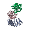
| ||||||||
|---|---|---|---|---|---|---|---|---|---|
| 1 |
| ||||||||
| Unit cell |
|
-Components
| #1: Protein | Mass: 25896.285 Da / Num. of mol.: 1 Source method: isolated from a genetically manipulated source Source: (gene. exp.) Homo sapiens (human) / Production host:  |
|---|---|
| #2: Protein | Mass: 35693.203 Da / Num. of mol.: 1 Source method: isolated from a genetically manipulated source Source: (gene. exp.) Homo sapiens (human) / Production host:   Spodoptera frugiperda (fall armyworm) / References: UniProt: P67775 Spodoptera frugiperda (fall armyworm) / References: UniProt: P67775 |
| #3: Protein | Mass: 34087.191 Da / Num. of mol.: 1 Source method: isolated from a genetically manipulated source Source: (gene. exp.) Homo sapiens (human) / Production host: |
| #4: Water | ChemComp-HOH /  Mass: 18.015 Da / Num. of mol.: 62 / Source method: isolated from a natural source / Formula: H2O Mass: 18.015 Da / Num. of mol.: 62 / Source method: isolated from a natural source / Formula: H2O |
| Sequence details | CHAIN A HAS 47-399 RESIDUES DELETION AND CHAIN P HAS 239-283 LOOP REPLACED BY EGK. THE PROTEIN ...CHAIN A HAS 47-399 RESIDUES DELETION AND CHAIN P HAS 239-283 LOOP REPLACED BY EGK. THE PROTEIN CONSTRUCT (CHAIN P) IS A NON-CATALYTIC MUTANT WITH SER156 REPLACED BY ALA. |
-Experimental details
-Experiment
| Experiment | Method: X-RAY DIFFRACTION / Number of used crystals: 1 |
|---|
- Sample preparation
| Crystal | Density Matthews: 2.17 Å3/Da / Density % sol: 43.22 % |
|---|---|
| Crystal grow | Temperature: 290 K / Method: vapor diffusion, hanging drop / pH: 7.5 Details: 25% w/v PEG3350, 100 mM ammonium citrate, and 5 mM DTT, pH 7.5, VAPOR DIFFUSION, HANGING DROP, temperature 290K |
-Data collection
| Diffraction | Mean temperature: 100 K |
|---|---|
| Diffraction source | Source: SYNCHROTRON / Site: NSLS  / Beamline: X29A / Wavelength: 1.0809 Å / Beamline: X29A / Wavelength: 1.0809 Å |
| Detector | Type: ADSC QUANTUM 315 / Detector: CCD / Date: Sep 10, 2007 |
| Radiation | Monochromator: Si(111) / Protocol: SINGLE WAVELENGTH / Monochromatic (M) / Laue (L): M / Scattering type: x-ray |
| Radiation wavelength | Wavelength: 1.0809 Å / Relative weight: 1 |
| Reflection | Resolution: 2.8→100 Å / Num. all: 20623 / Num. obs: 20623 / % possible obs: 98.6 % / Observed criterion σ(I): -3 / Redundancy: 3 % / Rsym value: 0.095 |
| Reflection shell | Resolution: 2.8→2.9 Å / Redundancy: 2.9 % / Rsym value: 0.4 / % possible all: 95.7 |
- Processing
| Software |
| ||||||||||||||||||||||||||||||||||||||||||||||||||||||||||||||||||||||||||||||||||||||||||||||||||||||||||||||||||||||||||||||||||||||||||||||||||||||||||||||||||||||||||
|---|---|---|---|---|---|---|---|---|---|---|---|---|---|---|---|---|---|---|---|---|---|---|---|---|---|---|---|---|---|---|---|---|---|---|---|---|---|---|---|---|---|---|---|---|---|---|---|---|---|---|---|---|---|---|---|---|---|---|---|---|---|---|---|---|---|---|---|---|---|---|---|---|---|---|---|---|---|---|---|---|---|---|---|---|---|---|---|---|---|---|---|---|---|---|---|---|---|---|---|---|---|---|---|---|---|---|---|---|---|---|---|---|---|---|---|---|---|---|---|---|---|---|---|---|---|---|---|---|---|---|---|---|---|---|---|---|---|---|---|---|---|---|---|---|---|---|---|---|---|---|---|---|---|---|---|---|---|---|---|---|---|---|---|---|---|---|---|---|---|---|---|
| Refinement | Method to determine structure: MOLECULAR REPLACEMENT Starting model: PP2A A subunit, PP2A C subunit, PME-1 monomer Resolution: 2.8→50 Å / Cor.coef. Fo:Fc: 0.933 / Cor.coef. Fo:Fc free: 0.874 / SU B: 34.63 / SU ML: 0.325 / TLS residual ADP flag: LIKELY RESIDUAL / Cross valid method: THROUGHOUT / σ(F): 0 / ESU R Free: 0.438 / Stereochemistry target values: MAXIMUM LIKELIHOOD
| ||||||||||||||||||||||||||||||||||||||||||||||||||||||||||||||||||||||||||||||||||||||||||||||||||||||||||||||||||||||||||||||||||||||||||||||||||||||||||||||||||||||||||
| Solvent computation | Ion probe radii: 0.8 Å / Shrinkage radii: 0.8 Å / VDW probe radii: 1.2 Å / Solvent model: BABINET MODEL WITH MASK | ||||||||||||||||||||||||||||||||||||||||||||||||||||||||||||||||||||||||||||||||||||||||||||||||||||||||||||||||||||||||||||||||||||||||||||||||||||||||||||||||||||||||||
| Displacement parameters | Biso mean: 44.744 Å2
| ||||||||||||||||||||||||||||||||||||||||||||||||||||||||||||||||||||||||||||||||||||||||||||||||||||||||||||||||||||||||||||||||||||||||||||||||||||||||||||||||||||||||||
| Refinement step | Cycle: LAST / Resolution: 2.8→50 Å
| ||||||||||||||||||||||||||||||||||||||||||||||||||||||||||||||||||||||||||||||||||||||||||||||||||||||||||||||||||||||||||||||||||||||||||||||||||||||||||||||||||||||||||
| Refine LS restraints |
| ||||||||||||||||||||||||||||||||||||||||||||||||||||||||||||||||||||||||||||||||||||||||||||||||||||||||||||||||||||||||||||||||||||||||||||||||||||||||||||||||||||||||||
| LS refinement shell | Resolution: 2.8→2.873 Å / Total num. of bins used: 20
| ||||||||||||||||||||||||||||||||||||||||||||||||||||||||||||||||||||||||||||||||||||||||||||||||||||||||||||||||||||||||||||||||||||||||||||||||||||||||||||||||||||||||||
| Refinement TLS params. | Method: refined / Refine-ID: X-RAY DIFFRACTION
| ||||||||||||||||||||||||||||||||||||||||||||||||||||||||||||||||||||||||||||||||||||||||||||||||||||||||||||||||||||||||||||||||||||||||||||||||||||||||||||||||||||||||||
| Refinement TLS group |
|
