 Movie
Movie Controller


+ Open data

- Basic information
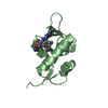
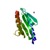
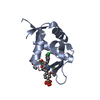
| Entry | Database: PDB / ID: 4rsv | ||||||
|---|---|---|---|---|---|---|---|
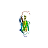
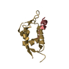
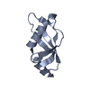
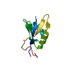
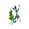
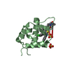
| Title | Human Obscurin Ig58 Domain | ||||||
 Components Components | Obscurin | ||||||
 Keywords Keywords | STRUCTURAL PROTEIN / Ig-like / cytoskeletal / Titin / Myocytes | ||||||
| Function / homology |  Function and homology information Function and homology informationprotein localization to M-band / phosphatidylinositol-5-phosphate binding / phosphatidylinositol-3-phosphate binding / phosphatidylinositol-3,4-bisphosphate binding / phosphatidylinositol-4-phosphate binding / M band / regulation of small GTPase mediated signal transduction / structural constituent of muscle / ankyrin binding / sarcomere organization ...protein localization to M-band / phosphatidylinositol-5-phosphate binding / phosphatidylinositol-3-phosphate binding / phosphatidylinositol-3,4-bisphosphate binding / phosphatidylinositol-4-phosphate binding / M band / regulation of small GTPase mediated signal transduction / structural constituent of muscle / ankyrin binding / sarcomere organization / NRAGE signals death through JNK / myofibril / RHOQ GTPase cycle / phosphatidylinositol-3,4,5-trisphosphate binding / RHOA GTPase cycle / titin binding / phosphatidylinositol-4,5-bisphosphate binding / guanyl-nucleotide exchange factor activity / sarcolemma / Z disc / G alpha (12/13) signalling events / calmodulin binding / non-specific serine/threonine protein kinase / nuclear body / protein serine kinase activity / protein serine/threonine kinase activity / ATP binding / metal ion binding / plasma membrane / cytosol Similarity search - Function | ||||||
| Biological species |  Homo sapiens (human) Homo sapiens (human) | ||||||
| Method |  X-RAY DIFFRACTION / SYNCHROTRON / MOLECULAR REPLACEMENT / Resolution: 2.41 Å X-RAY DIFFRACTION / SYNCHROTRON / MOLECULAR REPLACEMENT / Resolution: 2.41 Å | ||||||
 Authors Authors | Wright, N.T. / Oehler, M.C. / Berndsen, C.E. / Du Pont, K.E. | ||||||
 Citation Citation | Journal: To be Published Title: Arrhythmia and deregulated Ca2+ cycling underlie the development of hypertrophic cardiomyopathy due to mutant giant obscurin Authors: Hu, L.Y.R. / Ackermann, M.A. / Hecker, P.A. / Prosser, B.L. / King, B. / O'Connell, K.A. / Asico, L.D. / Jose, P.A. / Wright, N.T. / Oehler, M.C. / Berndsen, C.E. / Du Pont, K.E. / Meyer, L. ...Authors: Hu, L.Y.R. / Ackermann, M.A. / Hecker, P.A. / Prosser, B.L. / King, B. / O'Connell, K.A. / Asico, L.D. / Jose, P.A. / Wright, N.T. / Oehler, M.C. / Berndsen, C.E. / Du Pont, K.E. / Meyer, L.C. / Lederer, W.J. / Kontrogianni-Konstantopoulos, A. | ||||||
| History |
|
- Structure visualization
| Structure viewer | Molecule: MolmilJmol/JSmol |
|---|
- Downloads & links
-Download
| PDBx/mmCIF format | 4rsv.cif.gz | 29.8 KB | Display | PDBx/mmCIF format |
|---|---|---|---|---|
| PDB format | pdb4rsv.ent.gz | 20.1 KB | Display | PDB format |
| PDBx/mmJSON format | 4rsv.json.gz | Tree view | PDBx/mmJSON format | |
| Others |  Other downloads Other downloads |
-Validation report
| Summary document | 4rsv_validation.pdf.gz | 429.3 KB | Display | wwPDB validaton report |
|---|---|---|---|---|
| Full document | 4rsv_full_validation.pdf.gz | 434.2 KB | Display | |
| Data in XML | 4rsv_validation.xml.gz | 6.4 KB | Display | |
| Data in CIF | 4rsv_validation.cif.gz | 7.4 KB | Display | |
| Arichive directory | https://data.pdbj.org/pub/pdb/validation_reports/rs/4rsvftp://data.pdbj.org/pub/pdb/validation_reports/rs/4rsv | HTTPS FTP |
-Related structure data
| Related structure data |  2cr6S 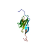 2e7bS  2rq8S 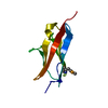 2yz8S S: Starting model for refinement |
|---|---|
| Similar structure data | |
| Other databases |


-Links
 PDBj
PDBj


- Assembly
| Deposited unit | 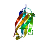
| ||||||||
|---|---|---|---|---|---|---|---|---|---|
| 1 |
| ||||||||
| Unit cell |
|
-Components
| #1: Protein | Mass: 11423.818 Da / Num. of mol.: 1 / Fragment: UNP residues 4337-4429 Source method: isolated from a genetically manipulated source Details: This sequence occurs naturally in humans / Source: (gene. exp.) Homo sapiens (human) / Gene: OBSCN, KIAA1556, KIAA1639 / Plasmid: PET24A / Production host:  References: UniProt: Q5VST9, non-specific serine/threonine protein kinase |
|---|---|
| #2: Water | ChemComp-HOH /  Mass: 18.015 Da / Num. of mol.: 1 / Source method: isolated from a natural source / Formula: H2O Mass: 18.015 Da / Num. of mol.: 1 / Source method: isolated from a natural source / Formula: H2O |
-Experimental details
-Experiment
| Experiment | Method: X-RAY DIFFRACTION / Number of used crystals: 2 |
|---|
- Sample preparation
| Crystal | Density Matthews: 2.22 Å3/Da / Density % sol: 44.68 % |
|---|---|
| Crystal grow | Temperature: 298 K / pH: 7.5 Details: 0.3-0.5M NaCl 19-23% PEG 3350 10.52-11.35 mg/ml, pH 7.5, VAPOR DIFFUSION, HANGING DROP, temperature 298K |
-Data collection
| Diffraction | Mean temperature: 170 K |
|---|---|
| Diffraction source | Source: SYNCHROTRON / Site: APS  / Beamline: 19-ID / Wavelength: 0.97918 / Beamline: 19-ID / Wavelength: 0.97918 |
| Detector | Type: ADSC QUANTUM 315r / Detector: CCD / Date: Apr 10, 2014 |
| Radiation | Monochromator: SI(111) / Protocol: SINGLE WAVELENGTH / Monochromatic (M) / Laue (L): M / Scattering type: x-ray |
| Radiation wavelength | Wavelength: 0.97918 Å / Relative weight: 1 |
| Reflection | Resolution: 2.411→37.64 Å / Num. obs: 4329 / % possible obs: 95.8 % / Observed criterion σ(I): 1 / Redundancy: 3.2 % / Rmerge(I) obs: 0.03344 / Rsym value: 0.04104 / Net I/σ(I): 29.47 |
| Reflection shell | Resolution: 2.56→2.62 Å / Redundancy: 3.4 % / Rmerge(I) obs: 0.3173 / Mean I/σ(I) obs: 3.08 / Rsym value: 0.04104 / % possible all: 69.2 |
- Processing
| Software |
| ||||||||||||||||||
|---|---|---|---|---|---|---|---|---|---|---|---|---|---|---|---|---|---|---|---|
| Refinement | Method to determine structure: MOLECULAR REPLACEMENT Starting model: 2CR6, 2YZ8, 2E7B, 2RQ8 Resolution: 2.41→37.64 Å / σ(F): 2 / Stereochemistry target values: ENGH & HUBER
| ||||||||||||||||||
| Refinement step | Cycle: LAST / Resolution: 2.41→37.64 Å
| ||||||||||||||||||
| Refine LS restraints |
|
