 Movie
Movie Controller

[English] 日本語
 Yorodumi
- PDB-2eff: Crystal structure analysis of the complex between CyaY and Co(II) -
+ Open data

- Basic information
| Entry | Database: PDB / ID: 2eff | ||||||
|---|---|---|---|---|---|---|---|
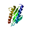
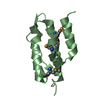
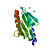
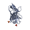
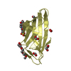
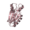
| Title | Crystal structure analysis of the complex between CyaY and Co(II) | ||||||
 Components Components | Protein cyaY | ||||||
 Keywords Keywords | UNKNOWN FUNCTION / FRATAXIN / FRIEDREICH'S ATAXIA IRON BINDING / IRON-SULFUR CLUSTER ASSEMBLY / DETOXIFYING REDOX-ACTIVE IRON | ||||||
| Function / homology |  Function and homology information Function and homology informationiron-sulfur cluster assembly / ferric iron binding / ferrous iron binding / cytosol / cytoplasm Similarity search - Function | ||||||
| Biological species |  | ||||||
| Method |  X-RAY DIFFRACTION / SYNCHROTRON / MOLECULAR REPLACEMENT / Resolution: 1.8 Å X-RAY DIFFRACTION / SYNCHROTRON / MOLECULAR REPLACEMENT / Resolution: 1.8 Å | ||||||
 Authors Authors | Sica, F. / Franzese, M. | ||||||
 Citation Citation | Journal: Febs J. / Year: 2007 Title: Understanding the binding properties of an unusual metal-binding protein - a study of bacterial frataxin Authors: Pastore, C. / Franzese, M. / Sica, F. / Temussi, P. / Pastore, A. #1: Journal: Structure / Year: 2004Title: Solution structure of the bacterial frataxin ortholog, CyaY: mapping the iron binding sites Authors: Nair, M. / Adinolfi, S. / Pastore, C. / Kelly, G. / Temussi, P. / Pastore, A. #2: Journal: Proc.Natl.Acad.Sci.USA / Year: 2000Title: Crystal structure of Escherichia coli CyaY protein reveals a previously unidentified fold for the evolutionarily conserved frataxin family Authors: Cho, S.J. / Lee, M.G. / Yang, J.K. / Lee, J.Y. / Song, H.K. / Suh, S.W. | ||||||
| History |
|
- Structure visualization
| Structure viewer | Molecule: MolmilJmol/JSmol |
|---|
- Downloads & links
-Download
| PDBx/mmCIF format | 2eff.cif.gz | 37.7 KB | Display | PDBx/mmCIF format |
|---|---|---|---|---|
| PDB format | pdb2eff.ent.gz | 24.7 KB | Display | PDB format |
| PDBx/mmJSON format | 2eff.json.gz | Tree view | PDBx/mmJSON format | |
| Others |  Other downloads Other downloads |
-Validation report
| Arichive directory | https://data.pdbj.org/pub/pdb/validation_reports/ef/2effftp://data.pdbj.org/pub/pdb/validation_reports/ef/2eff | HTTPS FTP |
|---|
-Related structure data
| Related structure data |  2p1xC  1ew4S S: Starting model for refinement C: citing same article ( |
|---|---|
| Similar structure data |


-Links
 PDBj
PDBj

- Assembly
| Deposited unit | 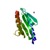
| ||||||||
|---|---|---|---|---|---|---|---|---|---|
| 1 |
| ||||||||
| Unit cell |
|
-Components
| #1: Protein | Mass: 12242.358 Da / Num. of mol.: 1 Source method: isolated from a genetically manipulated source Source: (gene. exp.) | ||
|---|---|---|---|
| #2: Chemical |   Mass: 58.933 Da / Num. of mol.: 2 / Source method: obtained synthetically / Formula: Co Mass: 58.933 Da / Num. of mol.: 2 / Source method: obtained synthetically / Formula: Co#3: Water | ChemComp-HOH / |  Mass: 18.015 Da / Num. of mol.: 135 / Source method: isolated from a natural source / Formula: H2O Mass: 18.015 Da / Num. of mol.: 135 / Source method: isolated from a natural source / Formula: H2O |
-Experimental details
-Experiment
| Experiment | Method: X-RAY DIFFRACTION / Number of used crystals: 1 |
|---|
- Sample preparation
| Crystal | Density Matthews: 2.23 Å3/Da / Density % sol: 44.91 % |
|---|---|
| Crystal grow | Temperature: 293 K / Method: vapor diffusion, sitting drop / pH: 4.7 Details: 30% w/v PEG 4000, 200mM CoCl2, 2mM beta-mercaptoethanol, 100 mM sodium acetate, pH 4.7, VAPOR DIFFUSION, SITTING DROP, temperature 293K |
-Data collection
| Diffraction | Mean temperature: 100 K |
|---|---|
| Diffraction source | Source: SYNCHROTRON / Site: ELETTRA  / Beamline: 5.2R / Wavelength: 1.2 Å / Beamline: 5.2R / Wavelength: 1.2 Å |
| Detector | Type: MAR CCD 130 mm / Detector: CCD / Date: Nov 8, 2005 |
| Radiation | Protocol: SINGLE WAVELENGTH / Monochromatic (M) / Laue (L): M / Scattering type: x-ray |
| Radiation wavelength | Wavelength: 1.2 Å / Relative weight: 1 |
| Reflection | Resolution: 1.75→30 Å / Num. obs: 20336 / % possible obs: 99.2 % / Redundancy: 5 % / Rmerge(I) obs: 0.057 / Net I/σ(I): 29.7 |
| Reflection shell | Resolution: 1.75→1.78 Å / Redundancy: 5 % / Rmerge(I) obs: 0.212 / Mean I/σ(I) obs: 6.9 / % possible all: 99.6 |
- Processing
| Software |
| |||||||||||||||||||||||||
|---|---|---|---|---|---|---|---|---|---|---|---|---|---|---|---|---|---|---|---|---|---|---|---|---|---|---|
| Refinement | Method to determine structure: MOLECULAR REPLACEMENT Starting model: PDB Entry 1EW4 Resolution: 1.8→18.6 Å / Isotropic thermal model: Isotropic / Cross valid method: THROUGHOUT / σ(F): 4 / Stereochemistry target values: Engh & Huber
| |||||||||||||||||||||||||
| Refinement step | Cycle: LAST / Resolution: 1.8→18.6 Å
| |||||||||||||||||||||||||
| Refine LS restraints |
|
