 Movie
Movie Controller


+ Open data

- Basic information
| Entry | Database: PDB / ID: 4d3g | ||||||
|---|---|---|---|---|---|---|---|
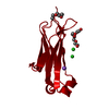
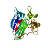
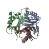
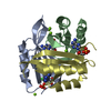
| Title | Structure of PstA | ||||||
 Components Components | PSTA | ||||||
 Keywords Keywords | SIGNALING PROTEIN / GRAM-POSITIVE / C-DI-AMP / PSTA | ||||||
| Function / homology |  Function and homology information Function and homology informationCyclic-di-AMP receptor / Cyclic-di-AMP receptor / Alpha-Beta Plaits - #120 / Nitrogen regulatory PII-like, alpha/beta / Nitrogen regulatory protein PII/ATP phosphoribosyltransferase, C-terminal / Alpha-Beta Plaits / 2-Layer Sandwich / Alpha Beta Similarity search - Domain/homology | ||||||
| Biological species |   STAPHYLOCOCCUS AUREUS (bacteria) STAPHYLOCOCCUS AUREUS (bacteria) | ||||||
| Method |  X-RAY DIFFRACTION / SYNCHROTRON / MOLECULAR REPLACEMENT / Resolution: 3 Å X-RAY DIFFRACTION / SYNCHROTRON / MOLECULAR REPLACEMENT / Resolution: 3 Å | ||||||
 Authors Authors | Campeotto, I. / Freemont, P. / Grundling, A. | ||||||
 Citation Citation | Journal: J.Biol.Chem. / Year: 2015 Title: Complex Structure and Biochemical Characterization of the Staphylococcus Aureus Cyclic Di-AMP Binding Protein Psta, the Founding Member of a New Signal Transduction Protein Family Authors: Campeotto, I. / Zhang, Y. / Mladenov, M.G. / Freemont, P.S. / Grundling, A. | ||||||
| History |
|
- Structure visualization
| Structure viewer | Molecule: MolmilJmol/JSmol |
|---|
- Downloads & links
-Download
| PDBx/mmCIF format | 4d3g.cif.gz | 45.4 KB | Display | PDBx/mmCIF format |
|---|---|---|---|---|
| PDB format | pdb4d3g.ent.gz | 31.9 KB | Display | PDB format |
| PDBx/mmJSON format | 4d3g.json.gz | Tree view | PDBx/mmJSON format | |
| Others |  Other downloads Other downloads |
-Validation report
| Arichive directory | https://data.pdbj.org/pub/pdb/validation_reports/d3/4d3gftp://data.pdbj.org/pub/pdb/validation_reports/d3/4d3g | HTTPS FTP |
|---|
-Related structure data
| Related structure data |  4d3hC 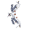 3m05S C: citing same article ( S: Starting model for refinement |
|---|---|
| Similar structure data |


-Links
 PDBj
PDBj- Assembly
| Deposited unit | 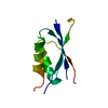
| ||||||||
|---|---|---|---|---|---|---|---|---|---|
| 1 | 
| ||||||||
| Unit cell |
|
-Components
| #1: Protein | Mass: 13886.558 Da / Num. of mol.: 1 Source method: isolated from a genetically manipulated source Source: (gene. exp.) STAPHYLOCOCCUS AUREUS (bacteria) / Production host: |
|---|---|
| Sequence details | 19 AA EXPRESSION |
-Experimental details
-Experiment
| Experiment | Method: X-RAY DIFFRACTION / Number of used crystals: 1 |
|---|
- Sample preparation
| Crystal | Density Matthews: 2.22 Å3/Da / Density % sol: 44.61 % / Description: NONE |
|---|---|
| Crystal grow | pH: 4.6 / Details: 200 MM NA MALONATE PH=4.6 AND 20% PEG3350 |
-Data collection
| Diffraction | Mean temperature: 100 K |
|---|---|
| Diffraction source | Source: SYNCHROTRON / Site: SOLEIL  / Beamline: PROXIMA 1 / Wavelength: 1.8233 / Beamline: PROXIMA 1 / Wavelength: 1.8233 |
| Detector | Type: DECTRIS PILATUS 6M / Detector: PIXEL / Date: Oct 2, 2013 |
| Radiation | Protocol: SINGLE WAVELENGTH / Monochromatic (M) / Laue (L): M / Scattering type: x-ray |
| Radiation wavelength | Wavelength: 1.8233 Å / Relative weight: 1 |
| Reflection | Resolution: 3→49.12 Å / Num. obs: 2672 / % possible obs: 100 % / Observed criterion σ(I): 7.9 / Redundancy: 38.2 % / Rmerge(I) obs: 0.11 / Net I/σ(I): 27.2 |
| Reflection shell | Resolution: 3→3.16 Å / Redundancy: 38.7 % / Rmerge(I) obs: 0.56 / Mean I/σ(I) obs: 7.9 / % possible all: 100 |
- Processing
| Software |
| ||||||||||||||||||||||||||||||||||||||||||||||||||||||||||||||||||||||||||||||||||||||||||||||||||||||||||||||||||||||||||||||||||||||||||||||||||||||||||||||||||||||||||||||||||||||
|---|---|---|---|---|---|---|---|---|---|---|---|---|---|---|---|---|---|---|---|---|---|---|---|---|---|---|---|---|---|---|---|---|---|---|---|---|---|---|---|---|---|---|---|---|---|---|---|---|---|---|---|---|---|---|---|---|---|---|---|---|---|---|---|---|---|---|---|---|---|---|---|---|---|---|---|---|---|---|---|---|---|---|---|---|---|---|---|---|---|---|---|---|---|---|---|---|---|---|---|---|---|---|---|---|---|---|---|---|---|---|---|---|---|---|---|---|---|---|---|---|---|---|---|---|---|---|---|---|---|---|---|---|---|---|---|---|---|---|---|---|---|---|---|---|---|---|---|---|---|---|---|---|---|---|---|---|---|---|---|---|---|---|---|---|---|---|---|---|---|---|---|---|---|---|---|---|---|---|---|---|---|---|---|
| Refinement | Method to determine structure: MOLECULAR REPLACEMENT Starting model: PDB ENTRY 3M05 Resolution: 3→49.18 Å / Cor.coef. Fo:Fc: 0.918 / Cor.coef. Fo:Fc free: 0.884 / SU B: 52.048 / SU ML: 0.401 / Cross valid method: THROUGHOUT / ESU R: 1.714 / ESU R Free: 0.437 / Stereochemistry target values: MAXIMUM LIKELIHOOD Details: HYDROGENS HAVE BEEN ADDED IN THE RIDING POSITIONS. U VALUES WITH TLS ADDED. DISORDERED REGIONS BETWEEN T33 AND N41 AND BETWEEN R67 AND V93 WERE NOT MODELLED
| ||||||||||||||||||||||||||||||||||||||||||||||||||||||||||||||||||||||||||||||||||||||||||||||||||||||||||||||||||||||||||||||||||||||||||||||||||||||||||||||||||||||||||||||||||||||
| Solvent computation | Ion probe radii: 0.8 Å / Shrinkage radii: 0.8 Å / VDW probe radii: 1.2 Å / Solvent model: MASK | ||||||||||||||||||||||||||||||||||||||||||||||||||||||||||||||||||||||||||||||||||||||||||||||||||||||||||||||||||||||||||||||||||||||||||||||||||||||||||||||||||||||||||||||||||||||
| Displacement parameters | Biso mean: 82.885 Å2
| ||||||||||||||||||||||||||||||||||||||||||||||||||||||||||||||||||||||||||||||||||||||||||||||||||||||||||||||||||||||||||||||||||||||||||||||||||||||||||||||||||||||||||||||||||||||
| Refinement step | Cycle: LAST / Resolution: 3→49.18 Å
| ||||||||||||||||||||||||||||||||||||||||||||||||||||||||||||||||||||||||||||||||||||||||||||||||||||||||||||||||||||||||||||||||||||||||||||||||||||||||||||||||||||||||||||||||||||||
| Refine LS restraints |
|