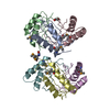
Deposited unit
A: Putative Pii-Like Signaling Protein
B: Putative Pii-Like Signaling Protein
C: Putative Pii-Like Signaling Protein
D: Putative Pii-Like Signaling Protein
E: Putative Pii-Like Signaling Protein
F: Putative Pii-Like Signaling Protein
hetero molecules Summary Component details
Theoretical mass Number of molelcules Total (without water) 73,038 8 Polymers 72,915 6 Non-polymers 122 2 Water 378 21
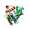
1
D: Putative Pii-Like Signaling Protein
E: Putative Pii-Like Signaling Protein
F: Putative Pii-Like Signaling Protein Summary Component details Symmetry operations Calculated values
Theoretical mass Number of molelcules Total (without water) 36,458 3 Polymers 36,458 3 Non-polymers 0 0 Water 54 3
Type Name Symmetry operation Number identity operation 1_555 x,y,z 1
Buried area 4030 Å2 ΔGint -18 kcal/mol Surface area 11030 Å2 Method
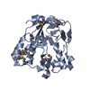
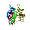
2
A: Putative Pii-Like Signaling Protein
B: Putative Pii-Like Signaling Protein
C: Putative Pii-Like Signaling Protein
hetero molecules Summary Component details Symmetry operations Calculated values
Theoretical mass Number of molelcules Total (without water) 36,580 5 Polymers 36,458 3 Non-polymers 122 2 Water 54 3
Type Name Symmetry operation Number identity operation 1_555 x,y,z 1
Buried area 4560 Å2 ΔGint -11 kcal/mol Surface area 10920 Å2 Method
Unit cell Length a, b, c (Å) 63.491, 78.749, 65.544 Angle α, β, γ (deg.) 90.000, 100.560, 90.000 Int Tables number 4 Space group name H-M P121 1
Noncrystallographic symmetry (NCS) NCS domain Show large table (3 x 48) Hide large table ID Ens-ID Details (eV)1 1 A2 1 B3 1 C4 1 D5 1 E6 1 F7 1 A8 1 B9 1 C10 1 D11 1 E12 1 F13 1 A14 1 B15 1 C16 1 D17 1 E18 1 F19 1 A20 1 B21 1 C22 1 D23 1 E24 1 F25 1 A26 1 B27 1 C28 1 D29 1 E30 1 F31 1 A32 1 B33 1 C34 1 D35 1 E36 1 F37 1 A38 1 B39 1 C40 1 D41 1 E42 1 F43 1 A44 1 B45 1 C46 1 D47 1 E48 1 F
NCS domain segments Ens-ID
Show large table (11 x 48) Hide large table Dom-ID Component-ID Beg auth comp-ID Beg label comp-ID End auth comp-ID End label comp-ID Refine code Auth asym-ID Label asym-ID Auth seq-ID Label seq-ID 1 1 LYSLYSGLYGLY2 AA3 - 31 4 - 32 2 1 LYSLYSGLYGLY2 BB3 - 31 4 - 32 3 1 LYSLYSGLYGLY2 CC3 - 31 4 - 32 4 1 LYSLYSGLYGLY2 DD3 - 31 4 - 32 5 1 LYSLYSGLYGLY2 EE3 - 31 4 - 32 6 1 LYSLYSGLYGLY2 FF3 - 31 4 - 32 7 2 TYRTYRTYRTYR5 AA32 33 8 2 TYRTYRTYRTYR5 BB32 33 9 2 TYRTYRTYRTYR5 CC32 33 10 2 TYRTYRTYRTYR5 DD32 33 11 2 TYRTYRTYRTYR5 EE32 33 12 2 TYRTYRTYRTYR5 FF32 33 13 3 THRTHRASPASP2 AA
 Movie
Movie Controller

 Yorodumi
 Open data
 Basic information
 Components
 Keywords
 Function and homology information
 Anabaena variabilis ATCC 29413 (bacteria)
 X-RAY DIFFRACTION /
  Authors
 Citation
 Structure visualization
 Downloads & links
 Other downloads
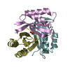


 PDBj
PDBj
 Assembly