 Movie
Movie Controller

[English] 日本語
 Yorodumi
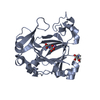
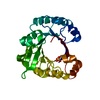
- PDB-4e22: Structure of cytidine monophosphate kinase from Yersinia pseudotu... -
+ Open data

- Basic information
| Entry | Database: PDB / ID: 4.0E+22 | ||||||
|---|---|---|---|---|---|---|---|
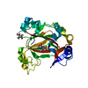
| Title | Structure of cytidine monophosphate kinase from Yersinia pseudotuberculosis | ||||||
 Components Components | Cytidylate kinase | ||||||
 Keywords Keywords | TRANSFERASE / P-loop / CMP/ATP binding | ||||||
| Function / homology |  Function and homology information Function and homology information(d)CMP kinase / CMP kinase activity / dCMP kinase activity / pyrimidine nucleotide metabolic process / nucleobase-containing small molecule interconversion / ATP binding / cytosol Similarity search - Function | ||||||
| Biological species |  Yersinia pseudotuberculosis (bacteria) Yersinia pseudotuberculosis (bacteria) | ||||||
| Method |  X-RAY DIFFRACTION / SYNCHROTRON / MOLECULAR REPLACEMENT / Resolution: 2.323 Å X-RAY DIFFRACTION / SYNCHROTRON / MOLECULAR REPLACEMENT / Resolution: 2.323 Å | ||||||
 Authors Authors | Clark, E.A. / Acharya, K.R. | ||||||
 Citation Citation | Journal: Open Biol / Year: 2012 Title: Structure and function of cytidine monophosphate kinase from Yersinia pseudotuberculosis, essential for virulence but not for survival. Authors: Walker, N.J. / Clark, E.A. / Ford, D.C. / Bullifent, H.L. / McAlister, E.V. / Duffield, M.L. / Acharya, K.R. / Oyston, P.C. | ||||||
| History |
|
- Structure visualization
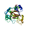
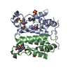
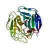
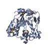
| Structure viewer | Molecule: MolmilJmol/JSmol |
|---|
- Downloads & links
-Download
| PDBx/mmCIF format | 4e22.cif.gz | 54.2 KB | Display | PDBx/mmCIF format |
|---|---|---|---|---|
| PDB format | pdb4e22.ent.gz | 38.5 KB | Display | PDB format |
| PDBx/mmJSON format | 4e22.json.gz | Tree view | PDBx/mmJSON format | |
| Others |  Other downloads Other downloads |
-Validation report
| Arichive directory | https://data.pdbj.org/pub/pdb/validation_reports/e2/4e22ftp://data.pdbj.org/pub/pdb/validation_reports/e2/4e22 | HTTPS FTP |
|---|
-Related structure data
| Similar structure data |
|---|
-Links
 PDBj
PDBj- Assembly
| Deposited unit | 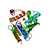
| ||||||||
|---|---|---|---|---|---|---|---|---|---|
| 1 |
| ||||||||
| Unit cell |
|
-Components
| #1: Protein | Mass: 27685.611 Da / Num. of mol.: 1 Source method: isolated from a genetically manipulated source Source: (gene. exp.) Yersinia pseudotuberculosis (bacteria) / Strain: YPIII / Gene: cmk, YPK_2669 / Production host: | ||
|---|---|---|---|
| #2: Chemical |   Mass: 96.063 Da / Num. of mol.: 2 / Source method: obtained synthetically / Formula: SO4 Mass: 96.063 Da / Num. of mol.: 2 / Source method: obtained synthetically / Formula: SO4#3: Water | ChemComp-HOH / |  Mass: 18.015 Da / Num. of mol.: 18 / Source method: isolated from a natural source / Formula: H2O Mass: 18.015 Da / Num. of mol.: 18 / Source method: isolated from a natural source / Formula: H2O |
-Experimental details
-Experiment
| Experiment | Method: X-RAY DIFFRACTION / Number of used crystals: 1 |
|---|
- Sample preparation
| Crystal | Density Matthews: 2.3 Å3/Da / Density % sol: 46.55 % |
|---|---|
| Crystal grow | Temperature: 289 K / Method: vapor diffusion, hanging drop / pH: 7.5 Details: 1 ul of 6 mg/ml Cmk (in 50 mM MES pH 6.0, 150 mM sodium chloride) plus 1 ul mother liquor (100 mM sodium HEPES pH 7.5, 1.2 M lithium sulfate, 100 mM magnesium chloride), VAPOR DIFFUSION, ...Details: 1 ul of 6 mg/ml Cmk (in 50 mM MES pH 6.0, 150 mM sodium chloride) plus 1 ul mother liquor (100 mM sodium HEPES pH 7.5, 1.2 M lithium sulfate, 100 mM magnesium chloride), VAPOR DIFFUSION, HANGING DROP, temperature 289K |
-Data collection
| Diffraction | Mean temperature: 100 K |
|---|---|
| Diffraction source | Source: SYNCHROTRON / Site: Diamond  / Beamline: I02 / Wavelength: 0.98 Å / Beamline: I02 / Wavelength: 0.98 Å |
| Detector | Type: ADSC QUANTUM 315 / Detector: CCD / Date: Apr 15, 2011 |
| Radiation | Monochromator: double crystal Si(III) / Protocol: SINGLE WAVELENGTH / Monochromatic (M) / Laue (L): M / Scattering type: x-ray |
| Radiation wavelength | Wavelength: 0.98 Å / Relative weight: 1 |
| Reflection | Resolution: 2.32→27.43 Å / Num. obs: 10266 / Redundancy: 4.3 % / Biso Wilson estimate: 61.6 Å2 / Rmerge(I) obs: 0.047 / Net I/σ(I): 16.2 |
- Processing
| Software |
| |||||||||||||||||||||||||||||||||||
|---|---|---|---|---|---|---|---|---|---|---|---|---|---|---|---|---|---|---|---|---|---|---|---|---|---|---|---|---|---|---|---|---|---|---|---|---|
| Refinement | Method to determine structure: MOLECULAR REPLACEMENT / Resolution: 2.323→27.43 Å / SU ML: 0.55 / σ(F): 1.97 / Phase error: 30.88 / Stereochemistry target values: ML
| |||||||||||||||||||||||||||||||||||
| Solvent computation | Shrinkage radii: 0.6 Å / VDW probe radii: 0.9 Å / Solvent model: FLAT BULK SOLVENT MODEL / Bsol: 53.479 Å2 / ksol: 0.378 e/Å3 | |||||||||||||||||||||||||||||||||||
| Displacement parameters |
| |||||||||||||||||||||||||||||||||||
| Refinement step | Cycle: LAST / Resolution: 2.323→27.43 Å
| |||||||||||||||||||||||||||||||||||
| Refine LS restraints |
| |||||||||||||||||||||||||||||||||||
| LS refinement shell |
|
