- PDB-4ril: Structure of the amyloid forming segment, GAVVTGVTAVA, from the N... -
+
データを開く
IDまたはキーワード:
読み込み中...
-
基本情報
登録情報
データベース: PDB / ID: 4ril
タイトル
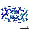
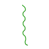
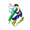
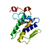
Structure of the amyloid forming segment, GAVVTGVTAVA, from the NAC domain of Parkinson's disease protein alpha-synuclein, residues 68-78, determined by electron diffraction
ジャーナル: Nature / 年: 2015 タイトル: Structure of the toxic core of α-synuclein from invisible crystals. 著者: Jose A Rodriguez / Magdalena I Ivanova / Michael R Sawaya / Duilio Cascio / Francis E Reyes / Dan Shi / Smriti Sangwan / Elizabeth L Guenther / Lisa M Johnson / Meng Zhang / Lin Jiang / Mark ...著者: Jose A Rodriguez / Magdalena I Ivanova / Michael R Sawaya / Duilio Cascio / Francis E Reyes / Dan Shi / Smriti Sangwan / Elizabeth L Guenther / Lisa M Johnson / Meng Zhang / Lin Jiang / Mark A Arbing / Brent L Nannenga / Johan Hattne / Julian Whitelegge / Aaron S Brewster / Marc Messerschmidt / Sébastien Boutet / Nicholas K Sauter / Tamir Gonen / David S Eisenberg / 要旨: The protein α-synuclein is the main component of Lewy bodies, the neuron-associated aggregates seen in Parkinson disease and other neurodegenerative pathologies. An 11-residue segment, which we term ...The protein α-synuclein is the main component of Lewy bodies, the neuron-associated aggregates seen in Parkinson disease and other neurodegenerative pathologies. An 11-residue segment, which we term NACore, appears to be responsible for amyloid formation and cytotoxicity of human α-synuclein. Here we describe crystals of NACore that have dimensions smaller than the wavelength of visible light and thus are invisible by optical microscopy. As the crystals are thousands of times too small for structure determination by synchrotron X-ray diffraction, we use micro-electron diffraction to determine the structure at atomic resolution. The 1.4 Å resolution structure demonstrates that this method can determine previously unknown protein structures and here yields, to our knowledge, the highest resolution achieved by any cryo-electron microscopy method to date. The structure exhibits protofibrils built of pairs of face-to-face β-sheets. X-ray fibre diffraction patterns show the similarity of NACore to toxic fibrils of full-length α-synuclein. The NACore structure, together with that of a second segment, inspires a model for most of the ordered portion of the toxic, full-length α-synuclein fibril, presenting opportunities for the design of inhibitors of α-synuclein fibrils.
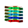
The biological unit is a pair of beta-sheets. One sheet is composed of chain A and unit cell translations along the b dimension. The other sheet is composed of the symmetry mate -x+1/2,y+1/2,-z, and unit cell translations along b.
-
要素
#1: タンパク質・ペプチド
Alpha-synuclein / Non-A beta component of AD amyloid / Non-A4 component of amyloid precursor / NACP
分子量: 944.083 Da / 分子数: 1 / 由来タイプ: 合成 詳細: Synthetic peptide GAVVTGVTAVA corresponding to segment 68-78 of human alpha-synuclein 由来: (合成) Homo sapiens (ヒト) / 参照: UniProt: P37840
名称: amyloid forming segment GAVVTGVTAVA from the NAC domain of alpha-synuclein タイプ: COMPLEX
試料
包埋: NO / シャドウイング: NO / 染色: NO / 凍結: YES
急速凍結
装置: FEI VITROBOT MARK IV / 凍結剤: ETHANE
結晶
マシュー密度: 1.46 Å3/Da / 溶媒含有率: 15.81 %
結晶化
温度: 310 K / 手法: batch crystallization / pH: 4 詳細: 1 mg of synthetic peptide GAVVTGVTAVA was dissolved in 1 ml of sterile water and shaken overnight in an orbital mixing plate, pH 4.0, batch crystallization, temperature 310K
-
データ収集
実験機器
モデル: Tecnai F20 / 画像提供: FEI Company
顕微鏡
モデル: FEI TECNAI F20
電子銃
加速電圧: 200 kV / 照射モード: FLOOD BEAM
電子レンズ
モード: DIFFRACTION
試料ホルダ
試料ホルダーモデル: GATAN 626 SINGLE TILT LIQUID NITROGEN CRYO TRANSFER HOLDER
撮影
平均露光時間: 3.5 sec. / 電子線照射量: 0.35 e/Å2 フィルム・検出器のモデル: TVIPS TEMCAM-F416 (4k x 4k)
回折
平均測定温度: 100 K
放射光源
由来: ELECTRON MICROSCOPE / タイプ: TECNAI F20 TEM / 波長: 0.0251 Å
 ムービー
ムービー コントローラー


 データを開く
 基本情報
基本情報 要素
要素 キーワード
キーワード 機能・相同性情報
 Homo sapiens (ヒト)
 分子置換 / クライオ電子顕微鏡法 / 解像度: 1.43 Å
  データ登録者
 引用
引用
 構造の表示
構造の表示 ダウンロードとリンク
 その他のダウンロード
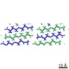


 PDBj
PDBj
 集合体
集合体
 分子量: 18.015 Da / 分子数: 2 / 由来タイプ: 天然 / 式: H2O
 試料調製
試料調製
 解析
解析