 Movie
Movie Controller

[English] 日本語
 Yorodumi
- PDB-4qvg: Crystal structure of S-adenosylmethionine-dependent methyltransfe... -
+ Open data

- Basic information
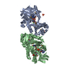
| Entry | Database: PDB / ID: 4qvg | ||||||
|---|---|---|---|---|---|---|---|
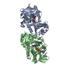
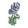
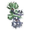
| Title | Crystal structure of S-adenosylmethionine-dependent methyltransferase SibL in its apo form | ||||||
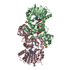
 Components Components | SibL | ||||||
 Keywords Keywords | TRANSFERASE / methyltransferase | ||||||
| Function / homology |  Function and homology information Function and homology informationS-adenosyl-L-methionine binding / O-methyltransferase activity / methyltransferase activity / methylation / protein dimerization activity Similarity search - Function | ||||||
| Biological species |  Streptosporangium sibiricum (bacteria) Streptosporangium sibiricum (bacteria) | ||||||
| Method |  X-RAY DIFFRACTION / SYNCHROTRON / MOLECULAR REPLACEMENT / Resolution: 2.9 Å X-RAY DIFFRACTION / SYNCHROTRON / MOLECULAR REPLACEMENT / Resolution: 2.9 Å | ||||||
 Authors Authors | Liu, J.S. / Chen, S.C. / Huang, C.H. / Yang, C.S. / Chen, Y. | ||||||
 Citation Citation | Journal: Sci Rep / Year: 2015 Title: Structure and mechanism of an antibiotics-synthesizing 3-hydroxykynurenine C-methyltransferase Authors: Chen, S.C. / Huang, C.H. / Lai, S.J. / Liu, J.S. / Fu, P.K. / Tseng, S.T. / Yang, C.S. / Lai, M.C. / Ko, T.P. / Chen, Y. | ||||||
| History |
|
- Structure visualization
| Structure viewer | Molecule: MolmilJmol/JSmol |
|---|
- Downloads & links
-Download
| PDBx/mmCIF format | 4qvg.cif.gz | 262 KB | Display | PDBx/mmCIF format |
|---|---|---|---|---|
| PDB format | pdb4qvg.ent.gz | 215.5 KB | Display | PDB format |
| PDBx/mmJSON format | 4qvg.json.gz | Tree view | PDBx/mmJSON format | |
| Others |  Other downloads Other downloads |
-Validation report
| Arichive directory | https://data.pdbj.org/pub/pdb/validation_reports/qv/4qvgftp://data.pdbj.org/pub/pdb/validation_reports/qv/4qvg | HTTPS FTP |
|---|
-Related structure data
| Related structure data | 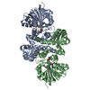 4u1qS S: Starting model for refinement |
|---|---|
| Similar structure data |
-Links
 PDBj
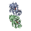
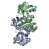
PDBj- Assembly
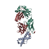
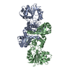
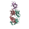
| Deposited unit | 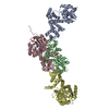
| ||||||||
|---|---|---|---|---|---|---|---|---|---|
| 1 | 
| ||||||||
| 2 | 
| ||||||||
| 3 | 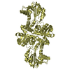
| ||||||||
| Unit cell |
|
-Components
| #1: Protein | Mass: 38923.797 Da / Num. of mol.: 4 Source method: isolated from a genetically manipulated source Source: (gene. exp.) Streptosporangium sibiricum (bacteria) / Gene: sibL / Production host: |
|---|
-Experimental details
-Experiment
| Experiment | Method: X-RAY DIFFRACTION / Number of used crystals: 1 |
|---|
- Sample preparation
| Crystal | Density Matthews: 3.93 Å3/Da / Density % sol: 68.73 % |
|---|---|
| Crystal grow | Temperature: 277 K / Method: vapor diffusion, sitting drop / pH: 9 Details: 10%(v/v) Polyethylene glycol 200, 0.1 M BIS-TRIS propane pH 9.0, 18%(w/v) Polyethylene glycol 8,000 , VAPOR DIFFUSION, SITTING DROP, temperature 277K |
-Data collection
| Diffraction | Mean temperature: 200 K |
|---|---|
| Diffraction source | Source: SYNCHROTRON / Site: NSRRC  / Beamline: BL13B1 / Wavelength: 0.9794 Å / Beamline: BL13B1 / Wavelength: 0.9794 Å |
| Detector | Type: ADSC QUANTUM 315r / Detector: CCD / Date: Jun 30, 2013 |
| Radiation | Monochromator: Si 111 CHANNEL / Protocol: SINGLE WAVELENGTH / Monochromatic (M) / Laue (L): M / Scattering type: x-ray |
| Radiation wavelength | Wavelength: 0.9794 Å / Relative weight: 1 |
| Reflection | Resolution: 2.9→30 Å / Num. all: 54894 / Num. obs: 54730 / % possible obs: 99.7 % / Observed criterion σ(F): 0 / Observed criterion σ(I): 0 |
| Reflection shell | Resolution: 2.9→3 Å / Redundancy: 5.8 % / Rmerge(I) obs: 0.211 / % possible all: 100 |
- Processing
| Software |
| ||||||||||||||||||||||||||||||||||||||||||||||||||||||||||||||||||||||||||||||||||||||||||
|---|---|---|---|---|---|---|---|---|---|---|---|---|---|---|---|---|---|---|---|---|---|---|---|---|---|---|---|---|---|---|---|---|---|---|---|---|---|---|---|---|---|---|---|---|---|---|---|---|---|---|---|---|---|---|---|---|---|---|---|---|---|---|---|---|---|---|---|---|---|---|---|---|---|---|---|---|---|---|---|---|---|---|---|---|---|---|---|---|---|---|---|
| Refinement | Method to determine structure: MOLECULAR REPLACEMENT Starting model: 4U1Q Resolution: 2.9→28.116 Å / SU ML: 0.36 / σ(F): 1.34 / Phase error: 29.04 / Stereochemistry target values: ML
| ||||||||||||||||||||||||||||||||||||||||||||||||||||||||||||||||||||||||||||||||||||||||||
| Solvent computation | Shrinkage radii: 0.9 Å / VDW probe radii: 1.11 Å / Solvent model: FLAT BULK SOLVENT MODEL | ||||||||||||||||||||||||||||||||||||||||||||||||||||||||||||||||||||||||||||||||||||||||||
| Refinement step | Cycle: LAST / Resolution: 2.9→28.116 Å
| ||||||||||||||||||||||||||||||||||||||||||||||||||||||||||||||||||||||||||||||||||||||||||
| Refine LS restraints |
| ||||||||||||||||||||||||||||||||||||||||||||||||||||||||||||||||||||||||||||||||||||||||||
| LS refinement shell | Refine-ID: X-RAY DIFFRACTION / Total num. of bins used: 14
|
