 Movie
Movie Controller

[English] 日本語
 Yorodumi
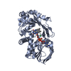
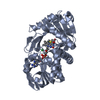
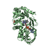
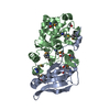
- PDB-3bhk: Crystal structure of R49K mutant of monomeric sarcosine oxidase c... -
+ Open data

- Basic information
| Entry | Database: PDB / ID: 3bhk | ||||||
|---|---|---|---|---|---|---|---|
| Title | Crystal structure of R49K mutant of monomeric sarcosine oxidase crystallized in phosphate as precipitant | ||||||
 Components Components | MONOMERIC SARCOSINE OXIDASE | ||||||
 Keywords Keywords | OXIDOREDUCTASE / FLAVOPROTEIN OXIDASE / FAD | ||||||
| Function / homology |  Function and homology information Function and homology informationsarcosine oxidase (formaldehyde-forming) / sarcosine oxidase activity / flavin adenine dinucleotide binding / cytoplasm Similarity search - Function | ||||||
| Biological species |  | ||||||
| Method |  X-RAY DIFFRACTION / SYNCHROTRON / Refined directly / Resolution: 1.71 Å X-RAY DIFFRACTION / SYNCHROTRON / Refined directly / Resolution: 1.71 Å | ||||||
 Authors Authors | Hassan-Abdallah, A. / Zhao, G. / Chen, Z. / Mathews, F.S. / Jorns, M.S. | ||||||
 Citation Citation | Journal: Biochemistry / Year: 2008 Title: Arginine 49 is a bifunctional residue important in catalysis and biosynthesis of monomeric sarcosine oxidase: a context-sensitive model for the electrostatic impact of arginine to lysine mutations. Authors: Hassan-Abdallah, A. / Zhao, G. / Chen, Z.W. / Mathews, F.S. / Schuman Jorns, M. #1: Journal: Structure / Year: 1999Title: Monomeric Sarcosine Oxidase: Structure of a Covalently Flavinylated Amine Oxidizing Enzyme Authors: Trickey, P. / Wagner, M.A. / Jorns, M.S. / Mathews, F.S. | ||||||
| History |
|
- Structure visualization
| Structure viewer | Molecule: MolmilJmol/JSmol |
|---|
- Downloads & links
-Download
| PDBx/mmCIF format | 3bhk.cif.gz | 180.4 KB | Display | PDBx/mmCIF format |
|---|---|---|---|---|
| PDB format | pdb3bhk.ent.gz | 139.9 KB | Display | PDB format |
| PDBx/mmJSON format | 3bhk.json.gz | Tree view | PDBx/mmJSON format | |
| Others |  Other downloads Other downloads |
-Validation report
| Arichive directory | https://data.pdbj.org/pub/pdb/validation_reports/bh/3bhkftp://data.pdbj.org/pub/pdb/validation_reports/bh/3bhk | HTTPS FTP |
|---|
-Related structure data
| Related structure data |  3bhfC  1l9f C: citing same article ( S: Starting model for refinement |
|---|---|
| Similar structure data |


-Links
 PDBj
PDBj- Assembly
| Deposited unit | 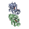
| ||||||||
|---|---|---|---|---|---|---|---|---|---|
| 1 | 
| ||||||||
| 2 | 
| ||||||||
| Unit cell |
|
-Components
| #1: Protein | Mass: 43204.535 Da / Num. of mol.: 2 / Mutation: R49K Source method: isolated from a genetically manipulated source Source: (gene. exp.) References: UniProt: P40859, sarcosine oxidase (formaldehyde-forming) #2: Chemical |   Mass: 35.453 Da / Num. of mol.: 2 / Source method: obtained synthetically / Formula: Cl Mass: 35.453 Da / Num. of mol.: 2 / Source method: obtained synthetically / Formula: Cl#3: Chemical |   Mass: 785.550 Da / Num. of mol.: 2 / Source method: obtained synthetically / Formula: C27H33N9O15P2 / Comment: FAD*YM Mass: 785.550 Da / Num. of mol.: 2 / Source method: obtained synthetically / Formula: C27H33N9O15P2 / Comment: FAD*YM#4: Chemical |   Mass: 92.094 Da / Num. of mol.: 2 / Source method: obtained synthetically / Formula: C3H8O3 Mass: 92.094 Da / Num. of mol.: 2 / Source method: obtained synthetically / Formula: C3H8O3#5: Water | ChemComp-HOH / |  Mass: 18.015 Da / Num. of mol.: 658 / Source method: isolated from a natural source / Formula: H2O Mass: 18.015 Da / Num. of mol.: 658 / Source method: isolated from a natural source / Formula: H2OHas protein modification | Y | |
|---|
-Experimental details
-Experiment
| Experiment | Method: X-RAY DIFFRACTION / Number of used crystals: 1 |
|---|
- Sample preparation
| Crystal | Density Matthews: 2.14 Å3/Da / Density % sol: 42.62 % |
|---|---|
| Crystal grow | Temperature: 295 K / Method: vapor diffusion, sitting drop / pH: 7 Details: 1.8 M Na/K phosphate, pH 7.0, VAPOR DIFFUSION, SITTING DROP, temperature 295K |
-Data collection
| Diffraction | Mean temperature: 100 K |
|---|---|
| Diffraction source | Source: SYNCHROTRON / Site: APS  / Beamline: 14-BM-C / Wavelength: 0.9 Å / Beamline: 14-BM-C / Wavelength: 0.9 Å |
| Detector | Type: ADSC QUANTUM 315 / Detector: CCD / Date: Apr 6, 2007 |
| Radiation | Protocol: SINGLE WAVELENGTH / Monochromatic (M) / Laue (L): M / Scattering type: x-ray |
| Radiation wavelength | Wavelength: 0.9 Å / Relative weight: 1 |
| Reflection | Resolution: 1.7→40 Å / Num. all: 78950 / Num. obs: 74292 / % possible obs: 94.1 % / Observed criterion σ(F): -0.6 / Observed criterion σ(I): -0.6 / Redundancy: 3.3 % / Biso Wilson estimate: 15.2 Å2 / Rmerge(I) obs: 0.063 / Net I/σ(I): 14.9 |
| Reflection shell | Resolution: 1.7→1.76 Å / Redundancy: 2.1 % / Rmerge(I) obs: 0.264 / Mean I/σ(I) obs: 2.3 / Num. unique all: 6441 / % possible all: 82.2 |
- Processing
| Software |
| ||||||||||||||||||||||||||||||||||||
|---|---|---|---|---|---|---|---|---|---|---|---|---|---|---|---|---|---|---|---|---|---|---|---|---|---|---|---|---|---|---|---|---|---|---|---|---|---|
| Refinement | Method to determine structure: Refined directly Starting model: PDB entry 1L9F 1l9f Resolution: 1.71→32.4 Å / Rfactor Rfree error: 0.003 / Data cutoff high absF: 386700.69 / Data cutoff low absF: 0 / Isotropic thermal model: RESTRAINED / Cross valid method: THROUGHOUT / σ(F): 0 / σ(I): 0 / Stereochemistry target values: Engh & Huber
| ||||||||||||||||||||||||||||||||||||
| Displacement parameters | Biso mean: 22.6 Å2 | ||||||||||||||||||||||||||||||||||||
| Refine analyze |
| ||||||||||||||||||||||||||||||||||||
| Refinement step | Cycle: LAST / Resolution: 1.71→32.4 Å
| ||||||||||||||||||||||||||||||||||||
| Refine LS restraints |
| ||||||||||||||||||||||||||||||||||||
| LS refinement shell | Resolution: 1.71→1.81 Å / Rfactor Rfree error: 0.013 / Total num. of bins used: 6
|
