 Movie
Movie Controller

[English] 日本語
 Yorodumi
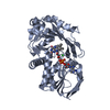
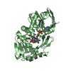
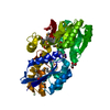
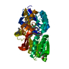
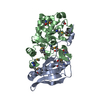
- PDB-3qse: Crystal structure for the complex of substrate-reduced msox with ... -
+ Open data

- Basic information
| Entry | Database: PDB / ID: 3qse | ||||||
|---|---|---|---|---|---|---|---|
| Title | Crystal structure for the complex of substrate-reduced msox with sarcosine | ||||||
 Components Components | Monomeric sarcosine oxidase | ||||||
 Keywords Keywords | OXIDOREDUCTASE / FLAVOPROTEIN / OXIDASE | ||||||
| Function / homology |  Function and homology information Function and homology informationsarcosine oxidase (formaldehyde-forming) / sarcosine oxidase activity / flavin adenine dinucleotide binding / cytoplasm Similarity search - Function | ||||||
| Biological species |  | ||||||
| Method |  X-RAY DIFFRACTION / SYNCHROTRON / MOLECULAR REPLACEMENT / Resolution: 1.75 Å X-RAY DIFFRACTION / SYNCHROTRON / MOLECULAR REPLACEMENT / Resolution: 1.75 Å | ||||||
 Authors Authors | Kommoju, P. / Chen, Z. / Bruckner, R.C. / Mathews, F.S. / Jorns, M.S. | ||||||
 Citation Citation | Journal: Biochemistry / Year: 2011 Title: Probing oxygen activation sites in two flavoprotein oxidases using chloride as an oxygen surrogate. Authors: Kommoju, P.R. / Chen, Z.W. / Bruckner, R.C. / Mathews, F.S. / Jorns, M.S. #1: Journal: Structure / Year: 1999Title: Monomeric sarcosine oxidase: structure of a covalently flavinylated amine oxidizing enzyme. Authors: Trickey, P. / Wagner, M.A. / Jorns, M.S. / Mathews, F.S. | ||||||
| History |
|
- Structure visualization
| Structure viewer | Molecule: MolmilJmol/JSmol |
|---|
- Downloads & links
-Download
| PDBx/mmCIF format | 3qse.cif.gz | 182.6 KB | Display | PDBx/mmCIF format |
|---|---|---|---|---|
| PDB format | pdb3qse.ent.gz | 141.4 KB | Display | PDB format |
| PDBx/mmJSON format | 3qse.json.gz | Tree view | PDBx/mmJSON format | |
| Others |  Other downloads Other downloads |
-Validation report
| Arichive directory | https://data.pdbj.org/pub/pdb/validation_reports/qs/3qseftp://data.pdbj.org/pub/pdb/validation_reports/qs/3qse | HTTPS FTP |
|---|
-Related structure data
| Related structure data |  3qsmC  3qssC  3qvpC 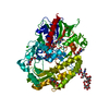 3qvrC  1l9f C: citing same article ( S: Starting model for refinement |
|---|---|
| Similar structure data |


-Links
 PDBj
PDBj- Assembly
| Deposited unit | 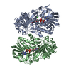
| ||||||||
|---|---|---|---|---|---|---|---|---|---|
| 1 | 
| ||||||||
| 2 | 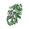
| ||||||||
| Unit cell |
|
-Components
| #1: Protein | Mass: 43101.355 Da / Num. of mol.: 2 Source method: isolated from a genetically manipulated source Source: (gene. exp.) References: UniProt: P40859, sarcosine oxidase (formaldehyde-forming) #2: Chemical |   Mass: 785.550 Da / Num. of mol.: 2 / Source method: obtained synthetically / Formula: C27H33N9O15P2 / Comment: FAD*YM Mass: 785.550 Da / Num. of mol.: 2 / Source method: obtained synthetically / Formula: C27H33N9O15P2 / Comment: FAD*YM#3: Chemical |   Mass: 35.453 Da / Num. of mol.: 2 / Source method: obtained synthetically / Formula: Cl Mass: 35.453 Da / Num. of mol.: 2 / Source method: obtained synthetically / Formula: Cl#4: Chemical |   Type: peptide linking / Mass: 89.093 Da / Num. of mol.: 2 / Source method: obtained synthetically / Formula: C3H7NO2 Type: peptide linking / Mass: 89.093 Da / Num. of mol.: 2 / Source method: obtained synthetically / Formula: C3H7NO2#5: Water | ChemComp-HOH / |  Mass: 18.015 Da / Num. of mol.: 774 / Source method: isolated from a natural source / Formula: H2O Mass: 18.015 Da / Num. of mol.: 774 / Source method: isolated from a natural source / Formula: H2OHas protein modification | Y | |
|---|
-Experimental details
-Experiment
| Experiment | Method: X-RAY DIFFRACTION / Number of used crystals: 1 |
|---|
- Sample preparation
| Crystal | Density Matthews: 2.1 Å3/Da / Density % sol: 41.37 % |
|---|---|
| Crystal grow | Temperature: 295 K / Method: vapor diffusion, hanging drop / pH: 7 Details: 1.9 M Na/K phosphate, 120 MM sarcosine, then soaked with 2.3 M NaCl, pH 7.0, VAPOR DIFFUSION, HANGING DROP, temperature 295K |
-Data collection
| Diffraction | Mean temperature: 100 K | |||||||||||||||||||||||||||||||||||||||||||||||||||||||||||||||||||||||||||||
|---|---|---|---|---|---|---|---|---|---|---|---|---|---|---|---|---|---|---|---|---|---|---|---|---|---|---|---|---|---|---|---|---|---|---|---|---|---|---|---|---|---|---|---|---|---|---|---|---|---|---|---|---|---|---|---|---|---|---|---|---|---|---|---|---|---|---|---|---|---|---|---|---|---|---|---|---|---|---|
| Diffraction source | Source: SYNCHROTRON / Site: APS  / Beamline: 14-BM-C / Wavelength: 0.9 Å / Beamline: 14-BM-C / Wavelength: 0.9 Å | |||||||||||||||||||||||||||||||||||||||||||||||||||||||||||||||||||||||||||||
| Detector | Type: ADSC QUANTUM 315 / Detector: CCD / Date: Dec 22, 2008 | |||||||||||||||||||||||||||||||||||||||||||||||||||||||||||||||||||||||||||||
| Radiation | Monochromator: APS Beamline 14-BM-C / Protocol: SINGLE WAVELENGTH / Monochromatic (M) / Laue (L): M / Scattering type: x-ray | |||||||||||||||||||||||||||||||||||||||||||||||||||||||||||||||||||||||||||||
| Radiation wavelength | Wavelength: 0.9 Å / Relative weight: 1 | |||||||||||||||||||||||||||||||||||||||||||||||||||||||||||||||||||||||||||||
| Reflection | Resolution: 1.75→40 Å / Num. all: 71893 / Num. obs: 70527 / % possible obs: 98.1 % / Observed criterion σ(F): -1 / Observed criterion σ(I): -1 / Redundancy: 4.6 % / Biso Wilson estimate: 16.5 Å2 / Rmerge(I) obs: 0.095 / Net I/σ(I): 14.1 | |||||||||||||||||||||||||||||||||||||||||||||||||||||||||||||||||||||||||||||
| Reflection shell |
|
- Processing
| Software |
| ||||||||||||||||||||||||||||||||||||
|---|---|---|---|---|---|---|---|---|---|---|---|---|---|---|---|---|---|---|---|---|---|---|---|---|---|---|---|---|---|---|---|---|---|---|---|---|---|
| Refinement | Method to determine structure: MOLECULAR REPLACEMENT Starting model: PDB entry 1L9F 1l9f Resolution: 1.75→25.19 Å / Rfactor Rfree error: 0.004 / Data cutoff high absF: 268939.76 / Data cutoff low absF: 0 / Isotropic thermal model: RESTRAINED / Cross valid method: THROUGHOUT / σ(F): 0 / σ(I): 0 / Stereochemistry target values: Engh & Huber
| ||||||||||||||||||||||||||||||||||||
| Solvent computation | Solvent model: FLAT MODEL / Bsol: 48.8423 Å2 / ksol: 0.35 e/Å3 | ||||||||||||||||||||||||||||||||||||
| Displacement parameters | Biso mean: 26.4 Å2
| ||||||||||||||||||||||||||||||||||||
| Refine analyze |
| ||||||||||||||||||||||||||||||||||||
| Refinement step | Cycle: LAST / Resolution: 1.75→25.19 Å
| ||||||||||||||||||||||||||||||||||||
| Refine LS restraints |
| ||||||||||||||||||||||||||||||||||||
| LS refinement shell | Resolution: 1.75→1.86 Å / Rfactor Rfree error: 0.02 / Total num. of bins used: 6
|
