- PDB-3dp7: CRYSTAL STRUCTURE OF SAM-dependent methyltransferase from Bactero... -
+
Open data
ID or keywords:
Loading...
-
Basic information
Entry
Database: PDB / ID: 3dp7
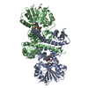
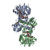
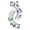
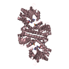
Title
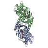
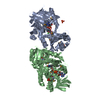
CRYSTAL STRUCTURE OF SAM-dependent methyltransferase from Bacteroides vulgatus ATCC 8482
Components
SAM-dependent methyltransferase
Keywords
TRANSFERASE / STRUCTURAL GENOMICS / PROTEIN STRUCTURE INITIATIVE / NEW YORK STRUCTURAL GENOMIX RESEARCH / SAM-dependent methyltransferase =CONSORTIUM / NYSGXRC / Methyltransferase / PSI-2 / New York SGX Research Center for Structural Genomics
Mass: 18.015 Da / Num. of mol.: 90 / Source method: isolated from a natural source / Formula: H2O
-
Experimental details
-
Experiment
Experiment
Method: X-RAY DIFFRACTION / Number of used crystals: 1
-
Sample preparation
Crystal
Density Matthews: 3.66 Å3/Da / Density % sol: 66.37 %
Crystal grow
Temperature: 294 K / Method: vapor diffusion, sitting drop / pH: 7 Details: 1.0 M sodium citrate, 0.1 M tris ph 7.0, 0.2 M nacl, VAPOR DIFFUSION, SITTING DROP, temperature 294K
Protocol: SINGLE WAVELENGTH / Scattering type: x-ray
Radiation wavelength
Wavelength: 0.979 Å / Relative weight: 1
Reflection
Redundancy: 4.2 % / Av σ(I) over netI: 10.9 / Number: 272651 / Rmerge(I) obs: 0.121 / Χ2: 5.32 / D res high: 2.33 Å / D res low: 50 Å / Num. obs: 65572 / % possible obs: 65.1
Diffraction reflection shell
Highest resolution (Å)
Lowest resolution (Å)
% possible obs (%)
ID
Rmerge(I) obs
Chi squared
Redundancy
5.02
50
100
1
0.091
12.671
4.7
3.98
5.02
100
1
0.121
9.128
4.7
3.48
3.98
100
1
0.135
4.371
4.8
3.16
3.48
100
1
0.197
1.857
4.7
2.94
3.16
98.5
1
0.332
0.946
4.3
2.76
2.94
77.9
1
0.398
0.81
3.1
2.62
2.76
48.8
1
0.446
0.711
2.1
2.51
2.62
18.6
1
0.626
0.783
1.5
2.41
2.51
5.6
1
0.748
1.2
2.33
2.41
1.8
1
0.595
1.1
Reflection
Resolution: 2.33→50 Å / Num. obs: 65572 / % possible obs: 65.1 % / Redundancy: 4.2 % / Rmerge(I) obs: 0.121 / Χ2: 5.323 / Net I/σ(I): 10.9
Reflection shell
Resolution (Å)
Redundancy (%)
Num. unique all
Χ2
Diffraction-ID
% possible all
Rmerge(I) obs
2.33-2.41
1.1
181
0.595
1
1.8
2.41-2.51
1.2
570
0.748
1
5.6
2.51-2.62
1.5
1875
0.783
1
18.6
0.626
2.62-2.76
2.1
4911
0.711
1
48.8
0.446
2.76-2.94
3.1
7844
0.81
1
77.9
0.398
2.94-3.16
4.3
9891
0.946
1
98.5
0.332
3.16-3.48
4.7
10079
1.857
1
100
0.197
3.48-3.98
4.8
10094
4.371
1
100
0.135
3.98-5.02
4.7
10051
9.128
1
100
0.121
5.02-50
4.7
10076
12.671
1
100
0.091
-
Phasing
Phasing
Method: SAD
-
Processing
Software
Name
Version
Classification
NB
DENZO
datareduction
SCALEPACK
datascaling
REFMAC
refinement
PDB_EXTRACT
3.006
dataextraction
HKL-2000
datareduction
PHENIX
phasing
Refinement
Method to determine structure: SAD / Resolution: 2.33→19.82 Å / Cor.coef. Fo:Fc: 0.959 / Cor.coef. Fo:Fc free: 0.917 / Occupancy max: 1 / Occupancy min: 0.5 / FOM work R set: 0.822 / SU B: 18.709 / SU ML: 0.206 / TLS residual ADP flag: LIKELY RESIDUAL / Cross valid method: THROUGHOUT / σ(F): 0 / ESU R: 0.356 / ESU R Free: 0.257 / Stereochemistry target values: MAXIMUM LIKELIHOOD / Details: HYDROGENS HAVE BEEN ADDED IN THE RIDING POSITIONS
Rfactor
Num. reflection
% reflection
Selection details
Rfree
0.253
1671
5.1 %
RANDOM
Rwork
0.183
-
-
-
obs
0.187
32966
64.95 %
-
Solvent computation
Ion probe radii: 0.8 Å / Shrinkage radii: 0.8 Å / VDW probe radii: 1.2 Å / Solvent model: BABINET MODEL WITH MASK
In the structure databanks used in Yorodumi, some data are registered as the other names, "COVID-19 virus" and "2019-nCoV". Here are the details of the virus and the list of structure data.
Jan 31, 2019. EMDB accession codes are about to change! (news from PDBe EMDB page)
EMDB accession codes are about to change! (news from PDBe EMDB page)
The allocation of 4 digits for EMDB accession codes will soon come to an end. Whilst these codes will remain in use, new EMDB accession codes will include an additional digit and will expand incrementally as the available range of codes is exhausted. The current 4-digit format prefixed with “EMD-” (i.e. EMD-XXXX) will advance to a 5-digit format (i.e. EMD-XXXXX), and so on. It is currently estimated that the 4-digit codes will be depleted around Spring 2019, at which point the 5-digit format will come into force.
The EM Navigator/Yorodumi systems omit the EMD- prefix.
Related info.:Q: What is EMD? / ID/Accession-code notation in Yorodumi/EM Navigator
Yorodumi is a browser for structure data from EMDB, PDB, SASBDB, etc.
This page is also the successor to EM Navigator detail page, and also detail information page/front-end page for Omokage search.
The word "yorodu" (or yorozu) is an old Japanese word meaning "ten thousand". "mi" (miru) is to see.
Related info.:EMDB / PDB / SASBDB / Comparison of 3 databanks / Yorodumi Search / Aug 31, 2016. New EM Navigator & Yorodumi / Yorodumi Papers / Jmol/JSmol / Function and homology information / Changes in new EM Navigator and Yorodumi
 Movie
Movie Controller

 Yorodumi
 Open data
 Basic information
 Components
 Keywords
 Function and homology information
 Bacteroides vulgatus (bacteria)
 X-RAY DIFFRACTION /
  Authors
 Citation
 Structure visualization
 Downloads & links
 Other downloads


 PDBj
PDBj Assembly
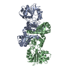
 Mass: 18.015 Da / Num. of mol.: 90 / Source method: isolated from a natural source / Formula: H2O
 Sample preparation
 / Beamline: 24-ID-C / Wavelength: 0.979 Å
 Processing