Resolution: 2.29→2.41 Å / Mean I/σ(I) obs: 5.3 / Rsym value: 0.187 / % possible all: 73.7
-
Processing
Software
Name
Version
Classification
MOSFLM
datareduction
SCALA
datascaling
PHASER
phasing
REFMAC
5.1.24
refinement
CCP4
(SCALA)
datascaling
Refinement
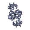
Method to determine structure: MOLECULAR REPLACEMENT Starting model: RdmB+SAM complex Resolution: 2.3→76 Å / Isotropic thermal model: Isotropic B-factors for each atom / Cross valid method: THROUGHOUT / Details: Hydrogens have been added in the riding position
Rfactor
Num. reflection
% reflection
Selection details
Rfree
0.29613
1910
-
RANDOM
Rwork
0.25102
-
-
-
obs
0.25324
36141
95.83 %
-
all
-
37903
-
-
Displacement parameters
Biso mean: 38.043 Å2
Baniso -1
Baniso -2
Baniso -3
1-
2.43 Å2
2.43 Å2
-3.65 Å2
2-
-
1.22 Å2
0 Å2
3-
-
-
0 Å2
Refinement step
Cycle: LAST / Resolution: 2.3→76 Å
Protein
Nucleic acid
Ligand
Solvent
Total
Num. atoms
5035
0
164
118
5317
Refine LS restraints
Refine-ID
Type
Dev ideal
X-RAY DIFFRACTION
r_bond_refined_d
0.013
X-RAY DIFFRACTION
r_angle_refined_deg
1.496
LS refinement shell
Resolution: 2.3→2.38 Å /
Rfactor
Num. reflection
Rfree
0.34
145
Rwork
0.3
-
obs
-
2383
+
About Yorodumi
-
News
-
Feb 9, 2022. New format data for meta-information of EMDB entries
New format data for meta-information of EMDB entries
Version 3 of the EMDB header file is now the official format.
The previous official version 1.9 will be removed from the archive.
In the structure databanks used in Yorodumi, some data are registered as the other names, "COVID-19 virus" and "2019-nCoV". Here are the details of the virus and the list of structure data.
Jan 31, 2019. EMDB accession codes are about to change! (news from PDBe EMDB page)
EMDB accession codes are about to change! (news from PDBe EMDB page)
The allocation of 4 digits for EMDB accession codes will soon come to an end. Whilst these codes will remain in use, new EMDB accession codes will include an additional digit and will expand incrementally as the available range of codes is exhausted. The current 4-digit format prefixed with “EMD-” (i.e. EMD-XXXX) will advance to a 5-digit format (i.e. EMD-XXXXX), and so on. It is currently estimated that the 4-digit codes will be depleted around Spring 2019, at which point the 5-digit format will come into force.
The EM Navigator/Yorodumi systems omit the EMD- prefix.
Related info.:Q: What is EMD? / ID/Accession-code notation in Yorodumi/EM Navigator
Yorodumi is a browser for structure data from EMDB, PDB, SASBDB, etc.
This page is also the successor to EM Navigator detail page, and also detail information page/front-end page for Omokage search.
The word "yorodu" (or yorozu) is an old Japanese word meaning "ten thousand". "mi" (miru) is to see.
Related info.:EMDB / PDB / SASBDB / Comparison of 3 databanks / Yorodumi Search / Aug 31, 2016. New EM Navigator & Yorodumi / Yorodumi Papers / Jmol/JSmol / Function and homology information / Changes in new EM Navigator and Yorodumi
 Movie
Movie Controller

 Yorodumi
 Open data
 Basic information
 Components
 Keywords
 Function and homology information
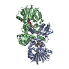
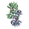
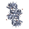
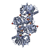
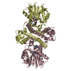
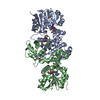
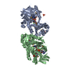
 Streptomyces purpurascens (bacteria)
 X-RAY DIFFRACTION /
  Authors
 Citation
 Structure visualization
 Downloads & links
 Other downloads


 PDBj
PDBj
 Assembly
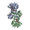

 Mass: 398.437 Da / Num. of mol.: 2 / Source method: obtained synthetically / Formula: C15H22N6O5S
 Mass: 769.831 Da / Num. of mol.: 2 / Source method: obtained synthetically / Formula: C40H51NO14
 Mass: 18.015 Da / Num. of mol.: 118 / Source method: isolated from a natural source / Formula: H2O
 Sample preparation
 / Beamline: ID14-1 / Wavelength: 0.934 Å
 Processing