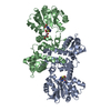
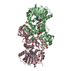
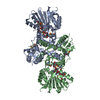
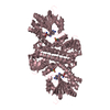
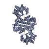
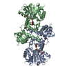
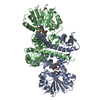
登録情報 データベース : PDB / ID : 3gxoタイトル Structure of the Mitomycin 7-O-methyltransferase MmcR with bound Mitomycin A MmcR キーワード / / / / 機能・相同性 分子機能 ドメイン・相同性 構成要素
/ / / / / / / / / / / / / / / / / / / / / / / / / / / / / / / / 生物種 Streptomyces lavendulae (バクテリア)手法 / / / 解像度 : 2.3 Å データ登録者 Singh, S. / Chang, A. / Bingman, C.A. / Phillips Jr., G.N. / Thorson, J.S. ジャーナル : Proteins / 年 : 2011タイトル : Structural characterization of the mitomycin 7-O-methyltransferase.著者 : Singh, S. / Chang, A. / Goff, R.D. / Bingman, C.A. / Gruschow, S. / Sherman, D.H. / Phillips, G.N. / Thorson, J.S. 履歴 登録 2009年4月2日 登録サイト / 処理サイト 改定 1.0 2010年4月21日 Provider / タイプ 改定 1.1 2011年7月13日 Group 改定 1.2 2017年11月1日 Group / カテゴリ 改定 1.3 2023年9月6日 Group Data collection / Database references ... Data collection / Database references / Derived calculations / Refinement description カテゴリ chem_comp_atom / chem_comp_bond ... chem_comp_atom / chem_comp_bond / database_2 / pdbx_initial_refinement_model / struct_ref_seq_dif / struct_site Item _database_2.pdbx_DOI / _database_2.pdbx_database_accession ... _database_2.pdbx_DOI / _database_2.pdbx_database_accession / _struct_ref_seq_dif.details / _struct_site.pdbx_auth_asym_id / _struct_site.pdbx_auth_comp_id / _struct_site.pdbx_auth_seq_id 改定 1.4 2023年11月22日 Group / カテゴリ / chem_comp_bond / Item / _chem_comp_bond.atom_id_2改定 1.5 2024年11月20日 Group / Structure summary / カテゴリ / pdbx_entry_details / Item / _chem_comp.type
すべて表示 表示を減らす
 ムービー
ムービー コントローラー


 データを開く
 基本情報
基本情報 要素
要素 キーワード
キーワード 機能・相同性情報
 Streptomyces lavendulae (バクテリア)
 X線回折 /
  データ登録者
 引用
引用 構造の表示
構造の表示 ダウンロードとリンク
 その他のダウンロード
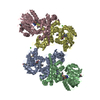


 PDBj
PDBj 集合体
集合体
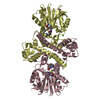

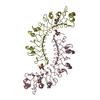
 分子量: 384.411 Da / 分子数: 4 / 由来タイプ: 合成 / 式: C14H20N6O5S
 分子量: 349.339 Da / 分子数: 4 / 由来タイプ: 合成 / 式: C16H19N3O6
 分子量: 40.078 Da / 分子数: 2 / 由来タイプ: 合成 / 式: Ca
 分子量: 18.015 Da / 分子数: 329 / 由来タイプ: 天然 / 式: H2O
 試料調製
試料調製 / ビームライン: 21-ID-F / 波長: 0.97872 Å
 解析
解析