- PDB-4qsz: Crystal structure of mouse JMJd7 fused with maltose-binding protein -
+
Open data
ID or keywords:
Loading...
-
Basic information
Entry
Database: PDB / ID: 4qsz
Title
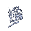
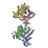
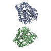
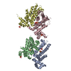
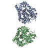
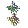
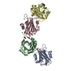
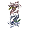
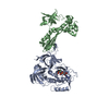
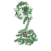
Crystal structure of mouse JMJd7 fused with maltose-binding protein
Components
Maltose-binding periplasmic protein, JmjC domain-containing protein 7 chimera
Keywords
TRANSCRIPTION / Demethylase
Function / homology
Function and homology information
peptidyl-lysine (3S)-dioxygenase / peptidyl-lysine 3-dioxygenase activity / protein hydroxylation / Protein hydroxylation / 2-oxoglutarate-dependent dioxygenase activity / Hydrolases; Acting on peptide bonds (peptidases) / detection of maltose stimulus / maltose transport complex / carbohydrate transport / aminopeptidase activity ...peptidyl-lysine (3S)-dioxygenase / peptidyl-lysine 3-dioxygenase activity / protein hydroxylation / Protein hydroxylation / 2-oxoglutarate-dependent dioxygenase activity / Hydrolases; Acting on peptide bonds (peptidases) / detection of maltose stimulus / maltose transport complex / carbohydrate transport / aminopeptidase activity / carbohydrate transmembrane transporter activity / maltose binding / maltose transport / maltodextrin transmembrane transport / ATP-binding cassette (ABC) transporter complex, substrate-binding subunit-containing / ATP-binding cassette (ABC) transporter complex / cell chemotaxis / monooxygenase activity / outer membrane-bounded periplasmic space / endopeptidase activity / periplasmic space / DNA damage response / proteolysis / membrane / metal ion binding / nucleus / cytoplasm Similarity search - Function
Cupin-like domain 8 / Cupin-like domain / A domain family that is part of the cupin metalloenzyme superfamily. / JmjC domain / JmjC domain profile. / Jelly Rolls / Maltose/Cyclodextrin ABC transporter, substrate-binding protein / Solute-binding family 1, conserved site / Bacterial extracellular solute-binding proteins, family 1 signature. / Bacterial extracellular solute-binding protein ...Cupin-like domain 8 / Cupin-like domain / A domain family that is part of the cupin metalloenzyme superfamily. / JmjC domain / JmjC domain profile. / Jelly Rolls / Maltose/Cyclodextrin ABC transporter, substrate-binding protein / Solute-binding family 1, conserved site / Bacterial extracellular solute-binding proteins, family 1 signature. / Bacterial extracellular solute-binding protein / Bacterial extracellular solute-binding protein / RmlC-like jelly roll fold / Jelly Rolls / Sandwich / Mainly Beta Similarity search - Domain/homology
CITRATE ANION / alpha-D-glucopyranose / Maltose/maltodextrin-binding periplasmic protein / Bifunctional peptidase and (3S)-lysyl hydroxylase Jmjd7 Similarity search - Component
Mass: 189.100 Da / Num. of mol.: 2 / Source method: obtained synthetically / Formula: C6H5O7
Has protein modification
Y
Sequence details
PROTEIN IS A CHIMERA COMPRISING RESIDUES 27-387 OF MBP (UNP P0AEX9) AND RESIDUES 2-312 OF JMJD7 ...PROTEIN IS A CHIMERA COMPRISING RESIDUES 27-387 OF MBP (UNP P0AEX9) AND RESIDUES 2-312 OF JMJD7 (UNP P0C872) CONNECTED BY AN AAAQTNAAAEF LINKER.
-
Experimental details
-
Experiment
Experiment
Method: X-RAY DIFFRACTION / Number of used crystals: 1
-
Sample preparation
Crystal
Density Matthews: 2.49 Å3/Da / Density % sol: 50.59 %
Crystal grow
Temperature: 277 K / Method: evaporation / pH: 8 Details: Hampton Research PEG/ION HT (89): 20 mM sodium citrate, 80 mM Bis-Tris propane, 16% PEG3350, pH 8.0, EVAPORATION, temperature 277K
-
Data collection
Diffraction
Mean temperature: 100 K
Diffraction source
Source: SYNCHROTRON / Site: ALS / Beamline: 4.2.2 / Wavelength: 1 Å
Detector
Type: NOIR-1 / Detector: CCD / Date: Mar 28, 2014
Radiation
Monochromator: Rosenbaum-Rock Si(111) sagitally focused / Protocol: SINGLE WAVELENGTH / Monochromatic (M) / Laue (L): M / Scattering type: x-ray
In the structure databanks used in Yorodumi, some data are registered as the other names, "COVID-19 virus" and "2019-nCoV". Here are the details of the virus and the list of structure data.
Jan 31, 2019. EMDB accession codes are about to change! (news from PDBe EMDB page)
EMDB accession codes are about to change! (news from PDBe EMDB page)
The allocation of 4 digits for EMDB accession codes will soon come to an end. Whilst these codes will remain in use, new EMDB accession codes will include an additional digit and will expand incrementally as the available range of codes is exhausted. The current 4-digit format prefixed with “EMD-” (i.e. EMD-XXXX) will advance to a 5-digit format (i.e. EMD-XXXXX), and so on. It is currently estimated that the 4-digit codes will be depleted around Spring 2019, at which point the 5-digit format will come into force.
The EM Navigator/Yorodumi systems omit the EMD- prefix.
Related info.:Q: What is EMD? / ID/Accession-code notation in Yorodumi/EM Navigator
Yorodumi is a browser for structure data from EMDB, PDB, SASBDB, etc.
This page is also the successor to EM Navigator detail page, and also detail information page/front-end page for Omokage search.
The word "yorodu" (or yorozu) is an old Japanese word meaning "ten thousand". "mi" (miru) is to see.
Related info.:EMDB / PDB / SASBDB / Comparison of 3 databanks / Yorodumi Search / Aug 31, 2016. New EM Navigator & Yorodumi / Yorodumi Papers / Jmol/JSmol / Function and homology information / Changes in new EM Navigator and Yorodumi
 Movie
Movie Controller

 Yorodumi
 Open data
 Basic information
 Components
 Keywords
 Function and homology information

 X-RAY DIFFRACTION /
  Authors
 Citation
 Structure visualization
 Downloads & links
 Other downloads


 PDBj
PDBj


 Assembly


 Type: D-saccharide, alpha linking / Mass: 180.156 Da / Num. of mol.: 4
 Mass: 189.100 Da / Num. of mol.: 2 / Source method: obtained synthetically / Formula: C6H5O7
 Sample preparation
 / Beamline: 4.2.2 / Wavelength: 1 Å
 Processing