 Movie
Movie Controller

[English] 日本語
 Yorodumi
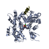
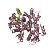
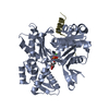
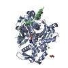
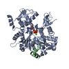
- PDB-3u9d: Crystal Structure of a chimera containing the N-terminal domain (... -
+ Open data

- Basic information
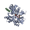
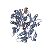
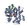
| Entry | Database: PDB / ID: 3u9d | ||||||
|---|---|---|---|---|---|---|---|
| Title | Crystal Structure of a chimera containing the N-terminal domain (residues 8-24) of drosophila Ciboulot and the C-terminal domain (residues 13-44) of bovine Thymosin-beta4, bound to G-actin-ATP | ||||||
 Components Components |
| ||||||
 Keywords Keywords | CONTRACTILE PROTEIN / PROTEIN BINDING | ||||||
| Function / homology |  Function and homology information Function and homology informationRegulation of CDH1 Function / actin monomer sequestering activity / Formation of the dystrophin-glycoprotein complex (DGC) / skeletal muscle fiber adaptation / Striated Muscle Contraction / larval central nervous system remodeling / positive regulation of neutrophil migration / negative regulation of actin filament polymerization / negative regulation of neutrophil chemotaxis / response to steroid hormone ...Regulation of CDH1 Function / actin monomer sequestering activity / Formation of the dystrophin-glycoprotein complex (DGC) / skeletal muscle fiber adaptation / Striated Muscle Contraction / larval central nervous system remodeling / positive regulation of neutrophil migration / negative regulation of actin filament polymerization / negative regulation of neutrophil chemotaxis / response to steroid hormone / cellular response to potassium ion / cellular response to glucocorticoid stimulus / positive regulation of wound healing / mesenchyme migration / striated muscle thin filament / skeletal muscle thin filament assembly / actin monomer binding / response to mechanical stimulus / skeletal muscle fiber development / stress fiber / regulation of cell migration / actin filament organization / sarcomere / filopodium / actin filament / protein sequestering activity / brain development / negative regulation of inflammatory response / Hydrolases; Acting on acid anhydrides; Acting on acid anhydrides to facilitate cellular and subcellular movement / lamellipodium / actin cytoskeleton / cell body / cytoskeleton / hydrolase activity / positive regulation of gene expression / : / ATP binding / cytosol / cytoplasm Similarity search - Function | ||||||
| Biological species |   | ||||||
| Method |  X-RAY DIFFRACTION / SYNCHROTRON / MOLECULAR REPLACEMENT / molecular replacement / Resolution: 2.5 Å X-RAY DIFFRACTION / SYNCHROTRON / MOLECULAR REPLACEMENT / molecular replacement / Resolution: 2.5 Å | ||||||
 Authors Authors | Renault, L. / Husson, C. / Carlier, M.F. / Didry, D. | ||||||
 Citation Citation | Journal: Embo J. / Year: 2012 Title: How a single residue in individual beta-thymosin/WH2 domains controls their functions in actin assembly. Authors: Didry, D. / Cantrelle, F.X. / Husson, C. / Roblin, P. / Moorthy, A.M. / Perez, J. / Le Clainche, C. / Hertzog, M. / Guittet, E. / Carlier, M.F. / van Heijenoort, C. / Renault, L. #1: Journal: Cell(Cambridge,Mass.) / Year: 2004Title: The beta-thymosin/WH2 domain; structural basis for the switch from inhibition to promotion of actin assembly. Authors: Hertzog, M. / van Heijenoort, C. / Didry, D. / Gaudier, M. / Coutant, J. / Gigant, B. / Didelot, G. / Preat, T. / Knossow, M. / Guittet, E. / Carlier, M.F. | ||||||
| History |
|
- Structure visualization
| Structure viewer | Molecule: MolmilJmol/JSmol |
|---|
- Downloads & links
-Download
| PDBx/mmCIF format | 3u9d.cif.gz | 304.9 KB | Display | PDBx/mmCIF format |
|---|---|---|---|---|
| PDB format | pdb3u9d.ent.gz | 246.7 KB | Display | PDB format |
| PDBx/mmJSON format | 3u9d.json.gz | Tree view | PDBx/mmJSON format | |
| Others |  Other downloads Other downloads |
-Validation report
| Arichive directory | https://data.pdbj.org/pub/pdb/validation_reports/u9/3u9dftp://data.pdbj.org/pub/pdb/validation_reports/u9/3u9d | HTTPS FTP |
|---|
-Related structure data
| Related structure data |  3sjhC  3u8xC  3u9zC 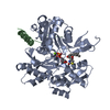 1sqkS C: citing same article ( S: Starting model for refinement |
|---|---|
| Similar structure data |


-Links
 PDBj
PDBj


- Assembly
| Deposited unit | 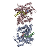
| ||||||||||||||||||||||||||||||||||||||||||||||||||||||||||||||||||||||||||||||||||||||||||||||||||||||
|---|---|---|---|---|---|---|---|---|---|---|---|---|---|---|---|---|---|---|---|---|---|---|---|---|---|---|---|---|---|---|---|---|---|---|---|---|---|---|---|---|---|---|---|---|---|---|---|---|---|---|---|---|---|---|---|---|---|---|---|---|---|---|---|---|---|---|---|---|---|---|---|---|---|---|---|---|---|---|---|---|---|---|---|---|---|---|---|---|---|---|---|---|---|---|---|---|---|---|---|---|---|---|---|
| 1 | 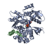
| ||||||||||||||||||||||||||||||||||||||||||||||||||||||||||||||||||||||||||||||||||||||||||||||||||||||
| 2 | 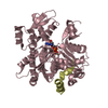
| ||||||||||||||||||||||||||||||||||||||||||||||||||||||||||||||||||||||||||||||||||||||||||||||||||||||
| Unit cell |
| ||||||||||||||||||||||||||||||||||||||||||||||||||||||||||||||||||||||||||||||||||||||||||||||||||||||
| Noncrystallographic symmetry (NCS) | NCS domain:
NCS domain segments:
NCS ensembles :
|
-Components
| #1: Protein | Mass: 41862.613 Da / Num. of mol.: 2 / Source method: isolated from a natural source / Details: organ: alpha Skeletal Muscle / Source: (natural) #2: Protein | Mass: 5962.708 Da / Num. of mol.: 2 Fragment: UNP O97428 residues 8-24, UNP P62326 residues 13-44 Source method: isolated from a genetically manipulated source Source: (gene. exp.) Gene: TMSB4, THYB4 / Plasmid: pGEX-6P1 / Production host:  #3: Chemical |   Mass: 507.181 Da / Num. of mol.: 2 / Source method: obtained synthetically / Formula: C10H16N5O13P3 / Comment: ATP, energy-carrying molecule*YM Mass: 507.181 Da / Num. of mol.: 2 / Source method: obtained synthetically / Formula: C10H16N5O13P3 / Comment: ATP, energy-carrying molecule*YM#4: Chemical |   Mass: 24.305 Da / Num. of mol.: 2 / Source method: obtained synthetically / Formula: Mg Mass: 24.305 Da / Num. of mol.: 2 / Source method: obtained synthetically / Formula: Mg |
|---|
-Experimental details
-Experiment
| Experiment | Method: X-RAY DIFFRACTION / Number of used crystals: 1 |
|---|
- Sample preparation
| Crystal | Density Matthews: 2.33 Å3/Da / Density % sol: 47.1 % |
|---|---|
| Crystal grow | Temperature: 298 K / Method: hanging drop / pH: 6.5 Details: 18% PEG3350, 0.05M NaAcetate pH4.7, 0.1M MgAcetate pH6.5, 0.32M Guanidine HCl, 0.8% Dioxane, hanging drop, temperature 298K |
-Data collection
| Diffraction | Mean temperature: 100 K | ||||||||||||||||||||||||||||||||||||||||||||||||||||||||||||||||||||||||||||||||||||||||||||||||||||||||||||||||||||||||
|---|---|---|---|---|---|---|---|---|---|---|---|---|---|---|---|---|---|---|---|---|---|---|---|---|---|---|---|---|---|---|---|---|---|---|---|---|---|---|---|---|---|---|---|---|---|---|---|---|---|---|---|---|---|---|---|---|---|---|---|---|---|---|---|---|---|---|---|---|---|---|---|---|---|---|---|---|---|---|---|---|---|---|---|---|---|---|---|---|---|---|---|---|---|---|---|---|---|---|---|---|---|---|---|---|---|---|---|---|---|---|---|---|---|---|---|---|---|---|---|---|---|
| Diffraction source | Source: SYNCHROTRON / Site: ESRF  / Beamline: ID29 / Wavelength: 0.97903 Å / Beamline: ID29 / Wavelength: 0.97903 Å | ||||||||||||||||||||||||||||||||||||||||||||||||||||||||||||||||||||||||||||||||||||||||||||||||||||||||||||||||||||||||
| Detector | Type: ADSC QUANTUM 315r / Detector: CCD / Date: Oct 5, 2006 | ||||||||||||||||||||||||||||||||||||||||||||||||||||||||||||||||||||||||||||||||||||||||||||||||||||||||||||||||||||||||
| Radiation | Protocol: SINGLE WAVELENGTH / Monochromatic (M) / Laue (L): M / Scattering type: x-ray | ||||||||||||||||||||||||||||||||||||||||||||||||||||||||||||||||||||||||||||||||||||||||||||||||||||||||||||||||||||||||
| Radiation wavelength | Wavelength: 0.97903 Å / Relative weight: 1 | ||||||||||||||||||||||||||||||||||||||||||||||||||||||||||||||||||||||||||||||||||||||||||||||||||||||||||||||||||||||||
| Reflection twin |
| ||||||||||||||||||||||||||||||||||||||||||||||||||||||||||||||||||||||||||||||||||||||||||||||||||||||||||||||||||||||||
| Reflection | Resolution: 2.5→20 Å / Num. obs: 29916 / % possible obs: 97.8 % / Observed criterion σ(I): -3 / Rmerge(I) obs: 0.061 / Net I/σ(I): 17.25 | ||||||||||||||||||||||||||||||||||||||||||||||||||||||||||||||||||||||||||||||||||||||||||||||||||||||||||||||||||||||||
| Reflection shell |
|
-Phasing
| Phasing | Method: molecular replacement | |||||||||
|---|---|---|---|---|---|---|---|---|---|---|
| Phasing MR | Model details: Phaser MODE: MR_AUTO
|
- Processing
| Software |
| ||||||||||||||||||||||||||||||||||||||||||||||||||||||||||||||||||||||||||||||||||||||||||||||||||||||||||||||||||||||||||||||||||||||||||||||||||||||||||||||||||||||||||
|---|---|---|---|---|---|---|---|---|---|---|---|---|---|---|---|---|---|---|---|---|---|---|---|---|---|---|---|---|---|---|---|---|---|---|---|---|---|---|---|---|---|---|---|---|---|---|---|---|---|---|---|---|---|---|---|---|---|---|---|---|---|---|---|---|---|---|---|---|---|---|---|---|---|---|---|---|---|---|---|---|---|---|---|---|---|---|---|---|---|---|---|---|---|---|---|---|---|---|---|---|---|---|---|---|---|---|---|---|---|---|---|---|---|---|---|---|---|---|---|---|---|---|---|---|---|---|---|---|---|---|---|---|---|---|---|---|---|---|---|---|---|---|---|---|---|---|---|---|---|---|---|---|---|---|---|---|---|---|---|---|---|---|---|---|---|---|---|---|---|---|---|
| Refinement | Method to determine structure: MOLECULAR REPLACEMENT Starting model: 1SQK Resolution: 2.5→19.64 Å / Cor.coef. Fo:Fc: 0.927 / Cor.coef. Fo:Fc free: 0.888 / Occupancy max: 1 / Occupancy min: 1 / SU B: 19.18 / SU ML: 0.194 / SU R Cruickshank DPI: 0.1256 / Cross valid method: THROUGHOUT / ESU R: 0.129 / ESU R Free: 0.06 / Stereochemistry target values: MAXIMUM LIKELIHOOD / Details: HYDROGENS HAVE BEEN ADDED IN THE RIDING POSITIONS
| ||||||||||||||||||||||||||||||||||||||||||||||||||||||||||||||||||||||||||||||||||||||||||||||||||||||||||||||||||||||||||||||||||||||||||||||||||||||||||||||||||||||||||
| Solvent computation | Ion probe radii: 0.8 Å / Shrinkage radii: 0.8 Å / VDW probe radii: 1.4 Å / Solvent model: MASK | ||||||||||||||||||||||||||||||||||||||||||||||||||||||||||||||||||||||||||||||||||||||||||||||||||||||||||||||||||||||||||||||||||||||||||||||||||||||||||||||||||||||||||
| Displacement parameters | Biso mean: 32.722 Å2
| ||||||||||||||||||||||||||||||||||||||||||||||||||||||||||||||||||||||||||||||||||||||||||||||||||||||||||||||||||||||||||||||||||||||||||||||||||||||||||||||||||||||||||
| Refinement step | Cycle: LAST / Resolution: 2.5→19.64 Å
| ||||||||||||||||||||||||||||||||||||||||||||||||||||||||||||||||||||||||||||||||||||||||||||||||||||||||||||||||||||||||||||||||||||||||||||||||||||||||||||||||||||||||||
| Refine LS restraints |
| ||||||||||||||||||||||||||||||||||||||||||||||||||||||||||||||||||||||||||||||||||||||||||||||||||||||||||||||||||||||||||||||||||||||||||||||||||||||||||||||||||||||||||
| Refine LS restraints NCS | Refine-ID: X-RAY DIFFRACTION
| ||||||||||||||||||||||||||||||||||||||||||||||||||||||||||||||||||||||||||||||||||||||||||||||||||||||||||||||||||||||||||||||||||||||||||||||||||||||||||||||||||||||||||
| LS refinement shell | Resolution: 2.498→2.562 Å / Total num. of bins used: 20
| ||||||||||||||||||||||||||||||||||||||||||||||||||||||||||||||||||||||||||||||||||||||||||||||||||||||||||||||||||||||||||||||||||||||||||||||||||||||||||||||||||||||||||
| Refinement TLS params. | Method: refined / Refine-ID: X-RAY DIFFRACTION
| ||||||||||||||||||||||||||||||||||||||||||||||||||||||||||||||||||||||||||||||||||||||||||||||||||||||||||||||||||||||||||||||||||||||||||||||||||||||||||||||||||||||||||
| Refinement TLS group |
|
