 Movie
Movie Controller

[English] 日本語
 Yorodumi
- PDB-3m1f: Crosslinked complex of actin with first W domain of Vibrio paraha... -
+ Open data

- Basic information
| Entry | Database: PDB / ID: 3m1f | ||||||
|---|---|---|---|---|---|---|---|
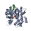
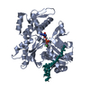
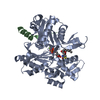
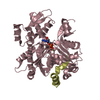
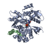
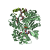
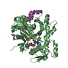
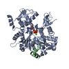
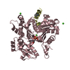
| Title | Crosslinked complex of actin with first W domain of Vibrio parahaemolyticus VopL | ||||||
 Components Components |
| ||||||
 Keywords Keywords | CONTRACTILE PROTEIN / ACTIN / ACTIN-BINDING PROTEIN / CROSSLINKING / NUCLEATOR / PROTEIN-PROTEIN INTERACTION / ATP-binding / Cytoskeleton / Methylation / Muscle protein / Nucleotide-binding / Phosphoprotein | ||||||
| Function / homology |  Function and homology information Function and homology informationcytoskeletal motor activator activity / myosin heavy chain binding / tropomyosin binding / actin filament bundle / troponin I binding / filamentous actin / mesenchyme migration / skeletal muscle myofibril / actin filament bundle assembly / striated muscle thin filament ...cytoskeletal motor activator activity / myosin heavy chain binding / tropomyosin binding / actin filament bundle / troponin I binding / filamentous actin / mesenchyme migration / skeletal muscle myofibril / actin filament bundle assembly / striated muscle thin filament / skeletal muscle thin filament assembly / actin monomer binding / skeletal muscle fiber development / stress fiber / titin binding / actin filament polymerization / filopodium / actin filament / Hydrolases; Acting on acid anhydrides; Acting on acid anhydrides to facilitate cellular and subcellular movement / calcium-dependent protein binding / lamellipodium / actin binding / cell body / protein domain specific binding / hydrolase activity / calcium ion binding / positive regulation of gene expression / magnesium ion binding / ATP binding / identical protein binding / cytoplasm Similarity search - Function | ||||||
| Biological species |    Vibrio parahaemolyticus (bacteria) Vibrio parahaemolyticus (bacteria) | ||||||
| Method |  X-RAY DIFFRACTION / SYNCHROTRON / MOLECULAR REPLACEMENT / Resolution: 2.89 Å X-RAY DIFFRACTION / SYNCHROTRON / MOLECULAR REPLACEMENT / Resolution: 2.89 Å | ||||||
 Authors Authors | Namgoong, S. / Dominguez, R. | ||||||
 Citation Citation | Journal: J.Mol.Biol. / Year: 2010 Title: Structure of a longitudinal actin dimer assembled by tandem w domains: implications for actin filament nucleation. Authors: Rebowski, G. / Namgoong, S. / Boczkowska, M. / Leavis, P.C. / Navaza, J. / Dominguez, R. | ||||||
| History |
|
- Structure visualization
| Structure viewer | Molecule: MolmilJmol/JSmol |
|---|
- Downloads & links
-Download
| PDBx/mmCIF format | 3m1f.cif.gz | 89.7 KB | Display | PDBx/mmCIF format |
|---|---|---|---|---|
| PDB format | pdb3m1f.ent.gz | 66.3 KB | Display | PDB format |
| PDBx/mmJSON format | 3m1f.json.gz | Tree view | PDBx/mmJSON format | |
| Others |  Other downloads Other downloads |
-Validation report
| Arichive directory | https://data.pdbj.org/pub/pdb/validation_reports/m1/3m1fftp://data.pdbj.org/pub/pdb/validation_reports/m1/3m1f | HTTPS FTP |
|---|
-Related structure data
| Related structure data | 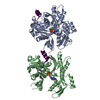 3m3nC 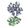 2a3zS S: Starting model for refinement C: citing same article ( |
|---|---|
| Similar structure data |

-Links
 PDBj
PDBj


- Assembly
| Deposited unit | 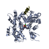
| ||||||||
|---|---|---|---|---|---|---|---|---|---|
| 1 |
| ||||||||
| Unit cell |
| ||||||||
| Details | The biological assembly is a monomer |
-Components
| #1: Protein | Mass: 41875.633 Da / Num. of mol.: 1 / Source method: isolated from a natural source / Source: (natural) |
|---|---|
| #2: Protein/peptide | Mass: 3426.897 Da / Num. of mol.: 1 / Fragment: VopL WH2 domain (130-160aa) / Mutation: V131C / Source method: obtained synthetically Details: Chemically synthesized peptide. The sequence naturally occurs in Vibrio parahaemolyticus. Source: (synth.) Vibrio parahaemolyticus (bacteria) / References: UniProt: Q87GE5 |
| #3: Chemical | ChemComp-ATP /   Mass: 507.181 Da / Num. of mol.: 1 / Source method: obtained synthetically / Formula: C10H16N5O13P3 / Comment: ATP, energy-carrying molecule*YM Mass: 507.181 Da / Num. of mol.: 1 / Source method: obtained synthetically / Formula: C10H16N5O13P3 / Comment: ATP, energy-carrying molecule*YM |
| #4: Chemical | ChemComp-CA /   Mass: 40.078 Da / Num. of mol.: 1 / Source method: obtained synthetically / Formula: Ca Mass: 40.078 Da / Num. of mol.: 1 / Source method: obtained synthetically / Formula: Ca |
-Experimental details
-Experiment
| Experiment | Method: X-RAY DIFFRACTION / Number of used crystals: 1 |
|---|
- Sample preparation
| Crystal | Density Matthews: 2.42 Å3/Da / Density % sol: 49.12 % |
|---|---|
| Crystal grow | Temperature: 298 K / Method: vapor diffusion, hanging drop / pH: 7.1 Details: 0.2 N Lithium Nitrate, 20% polyethylene glycol 3350, pH 7.1, VAPOR DIFFUSION, HANGING DROP, temperature 298.0K |
-Data collection
| Diffraction source | Source: SYNCHROTRON / Site: APS  / Beamline: 17-BM / Wavelength: 1 Å / Beamline: 17-BM / Wavelength: 1 Å |
|---|---|
| Detector | Type: MAR CCD 165 mm / Detector: CCD / Date: Oct 20, 2008 |
| Radiation | Monochromator: Si 111 CHANNEL / Protocol: SINGLE WAVELENGTH / Monochromatic (M) / Laue (L): M / Scattering type: x-ray |
| Radiation wavelength | Wavelength: 1 Å / Relative weight: 1 |
| Reflection | Resolution: 2.89→50 Å / Num. all: 18692 / Num. obs: 10207 / % possible obs: 99.2 % / Observed criterion σ(F): 2 / Observed criterion σ(I): 0 / Redundancy: 12.9 % / Biso Wilson estimate: 62.01 Å2 / Rmerge(I) obs: 0.168 / Rsym value: 0.168 / Net I/σ(I): 16.3 |
| Reflection shell | Resolution: 2.89→2.99 Å / Redundancy: 6.4 % / Rmerge(I) obs: 0.461 / Mean I/σ(I) obs: 1.8 / Rsym value: 0.461 / % possible all: 92.5 |
- Processing
| Software |
| |||||||||||||||||||||||||
|---|---|---|---|---|---|---|---|---|---|---|---|---|---|---|---|---|---|---|---|---|---|---|---|---|---|---|
| Refinement | Method to determine structure: MOLECULAR REPLACEMENT Starting model: 2a3z Resolution: 2.89→37.51 Å / Isotropic thermal model: Isotropic / Cross valid method: THROUGHOUT / σ(F): 0 / σ(I): 0 / Stereochemistry target values: Engh & Huber
| |||||||||||||||||||||||||
| Displacement parameters | Biso mean: 62.9 Å2 | |||||||||||||||||||||||||
| Refinement step | Cycle: LAST / Resolution: 2.89→37.51 Å
| |||||||||||||||||||||||||
| Refine LS restraints |
|
