 Movie
Movie Controller

[English] 日本語
 Yorodumi
- PDB-6jcu: Crystal structure of an actin monomer in complex with a nucleator... -
+ Open data

- Basic information
| Entry | Database: PDB / ID: 6jcu | ||||||
|---|---|---|---|---|---|---|---|
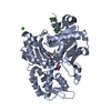
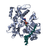
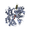
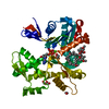
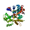
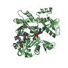
| Title | Crystal structure of an actin monomer in complex with a nucleator Cordon-Bleu WH2-motif peptide mutant. T22V, H11R | ||||||
 Components Components |
| ||||||
 Keywords Keywords | STRUCTURAL PROTEIN/PROTEIN BINDING / WH2 / actin / sequestering / PROTEIN BINDING / STRUCTURAL PROTEIN-PROTEIN BINDING complex | ||||||
| Function / homology |  Function and homology information Function and homology informationsomite specification / floor plate development / actin filament network formation / terminal web / embryonic axis specification / notochord development / actin crosslink formation / collateral sprouting in absence of injury / digestive tract development / positive regulation of dendrite development ...somite specification / floor plate development / actin filament network formation / terminal web / embryonic axis specification / notochord development / actin crosslink formation / collateral sprouting in absence of injury / digestive tract development / positive regulation of dendrite development / cytoskeletal motor activator activity / positive regulation of ruffle assembly / myosin heavy chain binding / dendritic growth cone / tropomyosin binding / actin filament bundle / troponin I binding / filamentous actin / mesenchyme migration / skeletal muscle myofibril / actin filament bundle assembly / striated muscle thin filament / skeletal muscle thin filament assembly / actin monomer binding / axonal growth cone / skeletal muscle fiber development / ruffle / stress fiber / titin binding / actin filament polymerization / liver development / neural tube closure / filopodium / actin filament / Hydrolases; Acting on acid anhydrides; Acting on acid anhydrides to facilitate cellular and subcellular movement / calcium-dependent protein binding / lamellipodium / actin cytoskeleton organization / cell body / cell cortex / protein domain specific binding / axon / hydrolase activity / neuronal cell body / calcium ion binding / positive regulation of gene expression / dendrite / perinuclear region of cytoplasm / magnesium ion binding / ATP binding / membrane / identical protein binding / plasma membrane / cytoplasm Similarity search - Function | ||||||
| Biological species |  | ||||||
| Method |  X-RAY DIFFRACTION / SYNCHROTRON / MOLECULAR REPLACEMENT / Resolution: 2.297 Å X-RAY DIFFRACTION / SYNCHROTRON / MOLECULAR REPLACEMENT / Resolution: 2.297 Å | ||||||
 Authors Authors | Scipion, C.P.M. / Robinson, R.C. | ||||||
 Citation Citation | Journal: To Be Published Title: Design of an actin-severing peptide. Authors: Scipion, C.P.M. / Robinson, R.C. | ||||||
| History |
|
- Structure visualization
| Structure viewer | Molecule: MolmilJmol/JSmol |
|---|
- Downloads & links
-Download
| PDBx/mmCIF format | 6jcu.cif.gz | 173.3 KB | Display | PDBx/mmCIF format |
|---|---|---|---|---|
| PDB format | pdb6jcu.ent.gz | 134.3 KB | Display | PDB format |
| PDBx/mmJSON format | 6jcu.json.gz | Tree view | PDBx/mmJSON format | |
| Others |  Other downloads Other downloads |
-Validation report
| Arichive directory | https://data.pdbj.org/pub/pdb/validation_reports/jc/6jcuftp://data.pdbj.org/pub/pdb/validation_reports/jc/6jcu | HTTPS FTP |
|---|
-Related structure data
| Related structure data | 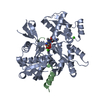 6jh8C 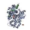 6jh9C 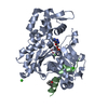 5ypuS S: Starting model for refinement C: citing same article ( |
|---|---|
| Similar structure data |


-Links
 PDBj
PDBj


- Assembly
| Deposited unit | 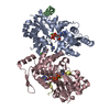
| ||||||||
|---|---|---|---|---|---|---|---|---|---|
| 1 | 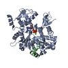
| ||||||||
| 2 | 
| ||||||||
| Unit cell |
| ||||||||
| Components on special symmetry positions |
|
-Components
| #1: Protein | Mass: 42109.973 Da / Num. of mol.: 2 Source method: isolated from a genetically manipulated source Source: (gene. exp.)  #2: Protein/peptide | Mass: 2431.859 Da / Num. of mol.: 2 / Mutation: H11R,T22V Source method: isolated from a genetically manipulated source Source: (gene. exp.) #3: Chemical |   Mass: 507.181 Da / Num. of mol.: 2 / Source method: obtained synthetically / Formula: C10H16N5O13P3 / Comment: ATP, energy-carrying molecule*YM Mass: 507.181 Da / Num. of mol.: 2 / Source method: obtained synthetically / Formula: C10H16N5O13P3 / Comment: ATP, energy-carrying molecule*YM#4: Chemical |   Mass: 40.078 Da / Num. of mol.: 3 / Source method: obtained synthetically / Formula: Ca Mass: 40.078 Da / Num. of mol.: 3 / Source method: obtained synthetically / Formula: Ca#5: Water | ChemComp-HOH / |  Mass: 18.015 Da / Num. of mol.: 281 / Source method: isolated from a natural source / Formula: H2O Mass: 18.015 Da / Num. of mol.: 281 / Source method: isolated from a natural source / Formula: H2O |
|---|
-Experimental details
-Experiment
| Experiment | Method: X-RAY DIFFRACTION / Number of used crystals: 1 |
|---|
- Sample preparation
| Crystal | Density Matthews: 2.28 Å3/Da / Density % sol: 46.06 % |
|---|---|
| Crystal grow | Temperature: 298.15 K / Method: vapor diffusion, sitting drop / Details: 0.2M Ammonium nitrate, 20% PEG 3350 |
-Data collection
| Diffraction | Mean temperature: 100 K / Serial crystal experiment: N |
|---|---|
| Diffraction source | Source: SYNCHROTRON / Site: NSRRC  / Beamline: TPS 05A / Wavelength: 1 Å / Beamline: TPS 05A / Wavelength: 1 Å |
| Detector | Type: RAYONIX MX300-HS / Detector: CCD / Date: Mar 29, 2017 |
| Radiation | Protocol: SINGLE WAVELENGTH / Monochromatic (M) / Laue (L): M / Scattering type: x-ray |
| Radiation wavelength | Wavelength: 1 Å / Relative weight: 1 |
| Reflection | Resolution: 2.297→92.106 Å / Num. obs: 835894 / % possible obs: 98.2 % / Redundancy: 6.7 % / Net I/σ(I): 14.6 |
| Reflection shell | Resolution: 2.297→2.364 Å |
- Processing
| Software |
| |||||||||||||||||||||||||||||||||||||||||||||||||||||||||||||||||||||||||||||||||||||||||||
|---|---|---|---|---|---|---|---|---|---|---|---|---|---|---|---|---|---|---|---|---|---|---|---|---|---|---|---|---|---|---|---|---|---|---|---|---|---|---|---|---|---|---|---|---|---|---|---|---|---|---|---|---|---|---|---|---|---|---|---|---|---|---|---|---|---|---|---|---|---|---|---|---|---|---|---|---|---|---|---|---|---|---|---|---|---|---|---|---|---|---|---|---|
| Refinement | Method to determine structure: MOLECULAR REPLACEMENT Starting model: 5YPU Resolution: 2.297→19.936 Å / SU ML: 0.25 / Cross valid method: FREE R-VALUE / σ(F): 1.36 / Phase error: 22.97
| |||||||||||||||||||||||||||||||||||||||||||||||||||||||||||||||||||||||||||||||||||||||||||
| Solvent computation | Shrinkage radii: 0.9 Å / VDW probe radii: 1.11 Å | |||||||||||||||||||||||||||||||||||||||||||||||||||||||||||||||||||||||||||||||||||||||||||
| Refinement step | Cycle: LAST / Resolution: 2.297→19.936 Å
| |||||||||||||||||||||||||||||||||||||||||||||||||||||||||||||||||||||||||||||||||||||||||||
| Refine LS restraints |
| |||||||||||||||||||||||||||||||||||||||||||||||||||||||||||||||||||||||||||||||||||||||||||
| LS refinement shell |
|
