 Movie
Movie Controller

[English] 日本語
 Yorodumi
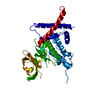
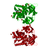
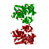
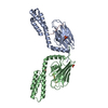
- PDB-3m3n: Structure of a Longitudinal Actin Dimer Assembled by Tandem W Domains -
+ Open data

- Basic information
| Entry | Database: PDB / ID: 3m3n | ||||||
|---|---|---|---|---|---|---|---|
| Title | Structure of a Longitudinal Actin Dimer Assembled by Tandem W Domains | ||||||
 Components Components |
| ||||||
 Keywords Keywords | STRUCTURAL PROTEIN / actin dimer / ATP-binding / actin cytoskeleton / methylation / muscle protein / actin-binding / MOTOR PROTEIN | ||||||
| Function / homology |  Function and homology information Function and homology informationactin monomer sequestering activity / negative regulation of membrane tubulation / spindle localization / membrane invagination / negative regulation of hematopoietic stem cell differentiation / plasma membrane tubulation / postsynaptic actin cytoskeleton organization / negative regulation of lymphocyte migration / actin nucleation / positive regulation of clathrin-dependent endocytosis ...actin monomer sequestering activity / negative regulation of membrane tubulation / spindle localization / membrane invagination / negative regulation of hematopoietic stem cell differentiation / plasma membrane tubulation / postsynaptic actin cytoskeleton organization / negative regulation of lymphocyte migration / actin nucleation / positive regulation of clathrin-dependent endocytosis / vesicle transport along actin filament / positive regulation of smooth muscle cell differentiation / regulation of cell projection assembly / actin cap / postsynapse organization / negative regulation of actin filament polymerization / Platelet degranulation / vesicle organization / vesicle budding from membrane / positive regulation of chemotaxis / dendritic spine morphogenesis / regulation of postsynapse organization / positive regulation of endothelial cell chemotaxis / positive regulation of filopodium assembly / protein-containing complex localization / positive regulation of smooth muscle cell migration / cytoskeletal motor activator activity / myosin heavy chain binding / tropomyosin binding / actin filament bundle / troponin I binding / filamentous actin / mesenchyme migration / cell leading edge / positive regulation of ATP biosynthetic process / skeletal muscle myofibril / actin filament bundle assembly / striated muscle thin filament / skeletal muscle thin filament assembly / actin monomer binding / positive regulation of blood vessel endothelial cell migration / skeletal muscle fiber development / cytoskeletal protein binding / stress fiber / titin binding / actin filament polymerization / regulation of cell migration / tumor necrosis factor-mediated signaling pathway / actin filament organization / negative regulation of canonical NF-kappaB signal transduction / response to bacterium / filopodium / actin filament / protein sequestering activity / negative regulation of inflammatory response / Hydrolases; Acting on acid anhydrides; Acting on acid anhydrides to facilitate cellular and subcellular movement / calcium-dependent protein binding / regulation of protein localization / lamellipodium / actin binding / actin cytoskeleton organization / cell body / cytoplasmic vesicle / cytoskeleton / postsynapse / Golgi membrane / protein domain specific binding / cell division / hydrolase activity / calcium ion binding / positive regulation of gene expression / positive regulation of DNA-templated transcription / glutamatergic synapse / magnesium ion binding / enzyme binding / positive regulation of transcription by RNA polymerase II / ATP binding / identical protein binding / nucleus / plasma membrane / cytoplasm / cytosol Similarity search - Function | ||||||
| Biological species |  | ||||||
| Method |  X-RAY DIFFRACTION / SYNCHROTRON / MOLECULAR REPLACEMENT / Resolution: 7 Å X-RAY DIFFRACTION / SYNCHROTRON / MOLECULAR REPLACEMENT / Resolution: 7 Å | ||||||
 Authors Authors | Rebowski, G. / Namgoong, S. / Dominguez, R. | ||||||
 Citation Citation | Journal: J.Mol.Biol. / Year: 2010 Title: Structure of a longitudinal actin dimer assembled by tandem w domains: implications for actin filament nucleation. Authors: Rebowski, G. / Namgoong, S. / Boczkowska, M. / Leavis, P.C. / Navaza, J. / Dominguez, R. | ||||||
| History |
|
- Structure visualization
| Structure viewer | Molecule: MolmilJmol/JSmol |
|---|
- Downloads & links
-Download
| PDBx/mmCIF format | 3m3n.cif.gz | 161.5 KB | Display | PDBx/mmCIF format |
|---|---|---|---|---|
| PDB format | pdb3m3n.ent.gz | 126.2 KB | Display | PDB format |
| PDBx/mmJSON format | 3m3n.json.gz | Tree view | PDBx/mmJSON format | |
| Others |  Other downloads Other downloads |
-Validation report
| Arichive directory | https://data.pdbj.org/pub/pdb/validation_reports/m3/3m3nftp://data.pdbj.org/pub/pdb/validation_reports/m3/3m3n | HTTPS FTP |
|---|
-Related structure data
| Related structure data |  3m1fSC S: Starting model for refinement C: citing same article ( |
|---|---|
| Similar structure data |


-Links
 PDBj
PDBj


- Assembly
| Deposited unit | 
| ||||||||
|---|---|---|---|---|---|---|---|---|---|
| 1 |
| ||||||||
| Unit cell |
|
-Components
| #1: Protein | Mass: 41875.633 Da / Num. of mol.: 2 / Source method: isolated from a natural source / Source: (natural) #2: Protein | | Mass: 11094.438 Da / Num. of mol.: 1 / Fragment: Engineered tandem W domain construct 3W Source method: isolated from a genetically manipulated source Source: (gene. exp.)  #3: Chemical |   Mass: 507.181 Da / Num. of mol.: 2 / Source method: obtained synthetically / Formula: C10H16N5O13P3 / Comment: ATP, energy-carrying molecule*YM Mass: 507.181 Da / Num. of mol.: 2 / Source method: obtained synthetically / Formula: C10H16N5O13P3 / Comment: ATP, energy-carrying molecule*YM#4: Chemical |   Mass: 40.078 Da / Num. of mol.: 2 / Source method: obtained synthetically / Formula: Ca Mass: 40.078 Da / Num. of mol.: 2 / Source method: obtained synthetically / Formula: Ca |
|---|
-Experimental details
-Experiment
| Experiment | Method: X-RAY DIFFRACTION / Number of used crystals: 1 |
|---|
- Sample preparation
| Crystal | Density Matthews: 3.54 Å3/Da / Density % sol: 65.29 % |
|---|---|
| Crystal grow | Temperature: 300 K / pH: 10 Details: 100 mM CAPS pH 10.0, and 24% PEG 3350, 100 mM RbCl, VAPOR DIFFUSION, HANGING DROP, temperature 300.0K |
-Data collection
| Diffraction | Mean temperature: 112 K |
|---|---|
| Diffraction source | Source: SYNCHROTRON / Site: APS  / Beamline: 17-BM / Wavelength: 1 / Beamline: 17-BM / Wavelength: 1 |
| Detector | Type: MAR CCD 165 mm / Detector: CCD / Date: Jul 23, 2009 Details: CYLINDRICALLY BENT ULE GLASS MIRROR WITH PT AND PD COATINGS |
| Radiation | Monochromator: CRYOGENICALLY-COOLED SI(111) DOUBLE-CRYSTAL SYSTEM Protocol: SINGLE WAVELENGTH / Monochromatic (M) / Laue (L): M / Scattering type: x-ray |
| Radiation wavelength | Wavelength: 1 Å / Relative weight: 1 |
| Reflection | Resolution: 7→50 Å / Num. obs: 2182 / % possible obs: 90 % / Redundancy: 20.5 % / Rmerge(I) obs: 0.086 / Net I/σ(I): 16.5 |
| Reflection shell | Resolution: 7→7.25 Å / Redundancy: 2.5 % / Rmerge(I) obs: 0.389 / Mean I/σ(I) obs: 1 / % possible all: 34.3 |
- Processing
| Software |
| ||||||||||||||
|---|---|---|---|---|---|---|---|---|---|---|---|---|---|---|---|
| Refinement | Method to determine structure: MOLECULAR REPLACEMENT Starting model: PDB ENTRY 3M1F Resolution: 7→50 Å / Details: NO REFINEMENT WAS PERFORMED | ||||||||||||||
| Refinement step | Cycle: LAST / Resolution: 7→50 Å
|
