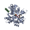
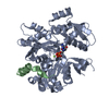
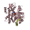
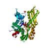
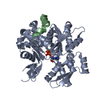
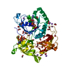
- PDB-3u9z: Crystal structure between actin and a protein construct containin... -
+
データを開く
IDまたはキーワード:
読み込み中...
-
基本情報
登録情報
データベース: PDB / ID: 3u9z
タイトル
Crystal structure between actin and a protein construct containing the first beta-thymosin domain of drosophila ciboulot (residues 2-58) with the three mutations N26D/Q27K/D28S
要素
Actin, alpha skeletal muscle
Ciboulot, isoform A
キーワード
CONTRACTILE PROTEIN / PROTEIN BINDING
機能・相同性
機能・相同性情報
larval central nervous system remodeling / cytoskeletal motor activator activity / myosin heavy chain binding / tropomyosin binding / actin filament bundle / troponin I binding / filamentous actin / mesenchyme migration / skeletal muscle myofibril / actin filament bundle assembly ...larval central nervous system remodeling / cytoskeletal motor activator activity / myosin heavy chain binding / tropomyosin binding / actin filament bundle / troponin I binding / filamentous actin / mesenchyme migration / skeletal muscle myofibril / actin filament bundle assembly / striated muscle thin filament / skeletal muscle thin filament assembly / actin monomer binding / skeletal muscle fiber development / stress fiber / titin binding / actin filament polymerization / actin filament organization / filopodium / actin filament / brain development / 加水分解酵素; 酸無水物に作用; 酸無水物に作用・細胞または細胞小器官の運動に関与 / calcium-dependent protein binding / lamellipodium / cell body / cytoskeleton / protein domain specific binding / hydrolase activity / calcium ion binding / positive regulation of gene expression / magnesium ion binding / ATP binding / identical protein binding / cytoplasm / cytosol 類似検索 - 分子機能
解像度: 2.09→46 Å / Cor.coef. Fo:Fc: 0.957 / Cor.coef. Fo:Fc free: 0.921 / WRfactor Rfree: 0.2194 / WRfactor Rwork: 0.1542 / Occupancy max: 1 / Occupancy min: 1 / FOM work R set: 0.8747 / SU B: 9.155 / SU ML: 0.112 / SU R Cruickshank DPI: 0.2054 / SU Rfree: 0.1868 / 交差検証法: THROUGHOUT / σ(F): 0 / ESU R Free: 0.187 / 立体化学のターゲット値: MAXIMUM LIKELIHOOD 詳細: HYDROGENS HAVE BEEN ADDED IN THE RIDING POSITIONS U VALUES : RESIDUAL ONLY
Rfactor
反射数
%反射
Selection details
Rfree
0.2266
1851
7.5 %
RANDOM
Rwork
0.1598
-
-
-
obs
0.1647
24652
99.93 %
-
all
-
24672
-
-
溶媒の処理
イオンプローブ半径: 0.8 Å / 減衰半径: 0.8 Å / VDWプローブ半径: 1.4 Å / 溶媒モデル: MASK
 ムービー
ムービー コントローラー


 データを開く
 基本情報
基本情報 要素
要素 キーワード
キーワード 機能・相同性情報

 X線回折 /
  データ登録者
 引用
引用 構造の表示
構造の表示 ダウンロードとリンク
 その他のダウンロード


 PDBj
PDBj


 集合体
集合体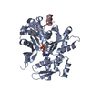


 分子量: 427.201 Da / 分子数: 1 / 由来タイプ: 合成 / 式: C10H15N5O10P2 / コメント: ADP, エネルギー貯蔵分子*YM
 分子量: 24.305 Da / 分子数: 1 / 由来タイプ: 合成 / 式: Mg
 分子量: 18.015 Da / 分子数: 368 / 由来タイプ: 天然 / 式: H2O
 試料調製
試料調製 / ビームライン: ID29 / 波長: 0.93 Å
 解析
解析