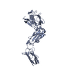
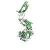
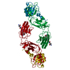
| Entry | Database: PDB / ID: 4qrb
|
|---|
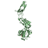
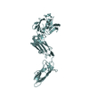
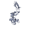
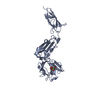
| Title | Structure and specificity of L-D-Transpeptidase from Mycobacterium tuberculosis and antibiotic resistance: Calcium binding promotes dimer formation |
|---|
 Components Components | L,d-transpeptidase LdtB |
|---|
 Keywords Keywords | HYDROLASE / Structural Genomics / Enzyme Function Initiative / Center for Structural Genomics of Infectious Diseases / CSGID / L-D-transpeptidase / D-D-transpeptidase / Single anomalous diffraction / imipenem / meropenem / peptidoglycan / beta-lactamase / peptide cross linkage / peptidoglycan stems / Bacterial cell wall periplasmic region |
|---|
| Function / homology |  Function and homology information Function and homology information
peptidoglycan-protein cross-linking / peptidoglycan L,D-transpeptidase activity / Transferases; Acyltransferases; Aminoacyltransferases / acyltransferase activity / cell wall organization / regulation of cell shape / metal ion binding / plasma membraneSimilarity search - Function Immunoglobulin-like - #3710 / Bacterial Ig domain, transpeptidase-associated / Bacterial Ig domain / L,D-transpeptidase catalytic domain-like / L,D-transpeptidase catalytic domain-like / : / L,D-transpeptidase (L,D-TPase) catalytic domain profile. / L,D-transpeptidase catalytic domain / L,D-transpeptidase catalytic domain-like / L,D-transpeptidase catalytic domain ...Immunoglobulin-like - #3710 / Bacterial Ig domain, transpeptidase-associated / Bacterial Ig domain / L,D-transpeptidase catalytic domain-like / L,D-transpeptidase catalytic domain-like / : / L,D-transpeptidase (L,D-TPase) catalytic domain profile. / L,D-transpeptidase catalytic domain / L,D-transpeptidase catalytic domain-like / L,D-transpeptidase catalytic domain / Prokaryotic membrane lipoprotein lipid attachment site profile. / Beta Barrel / Immunoglobulin-like / Sandwich / Mainly BetaSimilarity search - Domain/homology |
|---|
| Biological species |   Mycobacterium tuberculosis (bacteria) Mycobacterium tuberculosis (bacteria) |
|---|
| Method |  X-RAY DIFFRACTION / SYNCHROTRON / MOLECULAR REPLACEMENT / Resolution: 1.64 Å X-RAY DIFFRACTION / SYNCHROTRON / MOLECULAR REPLACEMENT / Resolution: 1.64 Å |
|---|
 Authors Authors | Gokulan, K. / Varughese, K.I. |
|---|
 Citation Citation | Journal: To be Published
Title: Structure and specificity of L-D-Transpeptidase from Mycobacterium tuberculosis and antibiotic resistance: Calcium binding promotes dimer formation
Authors: Gokulan, K. / Khare, S. / Cerniglia, C.E. / Foley, S.L. / Varughese, K.I. |
|---|
| History | | Deposition | Jun 30, 2014 | Deposition site: RCSB / Processing site: RCSB |
|---|
| Revision 1.0 | Dec 23, 2015 | Provider: repository / Type: Initial release |
|---|
| Revision 1.1 | Aug 24, 2016 | Group: Structure summary |
|---|
| Revision 1.2 | Feb 28, 2024 | Group: Data collection / Database references / Derived calculations
Category: chem_comp_atom / chem_comp_bond ...chem_comp_atom / chem_comp_bond / database_2 / struct_site
Item: _database_2.pdbx_DOI / _database_2.pdbx_database_accession ..._database_2.pdbx_DOI / _database_2.pdbx_database_accession / _struct_site.pdbx_auth_asym_id / _struct_site.pdbx_auth_comp_id / _struct_site.pdbx_auth_seq_id |
|---|
|
|---|
 Movie
Movie Controller

 Yorodumi
 Open data
 Basic information
 Structure visualization
 Downloads & links
 Other downloads


 PDBj
PDBj


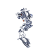
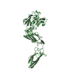
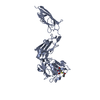
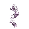
 Assembly
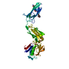
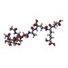
 Mass: 921.899 Da / Num. of mol.: 1 / Source method: obtained synthetically / Formula: C37H59N7O20
 Mass: 18.015 Da / Num. of mol.: 209 / Source method: isolated from a natural source / Formula: H2O
 Sample preparation
 / Beamline: BL9-2
 Processing