 Movie
Movie Controller


+ Open data

- Basic information
| Entry | Database: PDB / ID: 5du7 | ||||||
|---|---|---|---|---|---|---|---|
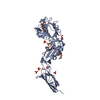
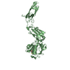
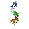
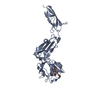
| Title | Crystal structure of ldtMt2 at 1.79 Angstrom resolution | ||||||
 Components Components | L,D-transpeptidase 2 | ||||||
 Keywords Keywords | TRANSFERASE / Peptidoglycan synthesis enzyme / cell wall enzyme | ||||||
| Function / homology |  Function and homology information Function and homology informationpeptidoglycan-based cell wall biogenesis / peptidoglycan-protein cross-linking / peptidoglycan metabolic process / peptidoglycan L,D-transpeptidase activity / Transferases; Acyltransferases; Aminoacyltransferases / acyltransferase activity / peptidoglycan-based cell wall / cell wall organization / regulation of cell shape / extracellular region ...peptidoglycan-based cell wall biogenesis / peptidoglycan-protein cross-linking / peptidoglycan metabolic process / peptidoglycan L,D-transpeptidase activity / Transferases; Acyltransferases; Aminoacyltransferases / acyltransferase activity / peptidoglycan-based cell wall / cell wall organization / regulation of cell shape / extracellular region / metal ion binding / plasma membrane Similarity search - Function | ||||||
| Biological species |   Mycobacterium tuberculosis (bacteria) Mycobacterium tuberculosis (bacteria) | ||||||
| Method |  X-RAY DIFFRACTION / SYNCHROTRON / MOLECULAR REPLACEMENT / molecular replacement / Resolution: 1.79 Å X-RAY DIFFRACTION / SYNCHROTRON / MOLECULAR REPLACEMENT / molecular replacement / Resolution: 1.79 Å | ||||||
 Authors Authors | Kumar, P. / Lamichhane, G. | ||||||
| Funding support |  United States, 1items United States, 1items
| ||||||
 Citation Citation | Journal: Nat. Chem. Biol. / Year: 2017 Title: Non-classical transpeptidases yield insight into new antibacterials. Authors: Kumar, P. / Kaushik, A. / Lloyd, E.P. / Li, S.G. / Mattoo, R. / Ammerman, N.C. / Bell, D.T. / Perryman, A.L. / Zandi, T.A. / Ekins, S. / Ginell, S.L. / Townsend, C.A. / Freundlich, J.S. / Lamichhane, G. | ||||||
| History |
|
- Structure visualization
| Structure viewer | Molecule: MolmilJmol/JSmol |
|---|
- Downloads & links
-Download
| PDBx/mmCIF format | 5du7.cif.gz | 314.9 KB | Display | PDBx/mmCIF format |
|---|---|---|---|---|
| PDB format | pdb5du7.ent.gz | 250.5 KB | Display | PDB format |
| PDBx/mmJSON format | 5du7.json.gz | Tree view | PDBx/mmJSON format | |
| Others |  Other downloads Other downloads |
-Validation report
| Arichive directory | https://data.pdbj.org/pub/pdb/validation_reports/du/5du7ftp://data.pdbj.org/pub/pdb/validation_reports/du/5du7 | HTTPS FTP |
|---|
-Related structure data
| Related structure data |  5dujC  5dvpC  5dzjC  5dzpC  5e1gC  5e1iC  5e51C  5e5lC  5k69C  3vynS C: citing same article ( S: Starting model for refinement |
|---|---|
| Similar structure data |


-Links
 PDBj
PDBj


- Assembly
| Deposited unit | 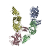
| ||||||||
|---|---|---|---|---|---|---|---|---|---|
| 1 | 
| ||||||||
| 2 | 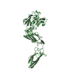
| ||||||||
| 3 | 
| ||||||||
| 4 | 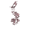
| ||||||||
| 5 | 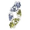
| ||||||||
| 6 | 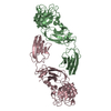
| ||||||||
| Unit cell |
|
-Components
| #1: Protein | Mass: 39532.004 Da / Num. of mol.: 4 / Fragment: residues 42-408 Source method: isolated from a genetically manipulated source Source: (gene. exp.) Mycobacterium tuberculosis (bacteria) / Strain: CDC 1551 / Oshkosh / Gene: ldtB, MT2594, V735_02606 / Plasmid: pET28a / Production host: References: UniProt: O53223, UniProt: I6Y9J2*PLUS, Transferases; Acyltransferases; Aminoacyltransferases #2: Water | ChemComp-HOH / |  Mass: 18.015 Da / Num. of mol.: 1827 / Source method: isolated from a natural source / Formula: H2O Mass: 18.015 Da / Num. of mol.: 1827 / Source method: isolated from a natural source / Formula: H2O |
|---|
-Experimental details
-Experiment
| Experiment | Method: X-RAY DIFFRACTION / Number of used crystals: 1 |
|---|
- Sample preparation
| Crystal | Density Matthews: 2.91 Å3/Da / Density % sol: 57.73 % |
|---|---|
| Crystal grow | Temperature: 291 K / Method: vapor diffusion, hanging drop / pH: 7.5 / Details: 20% PEG5000MME, 0.2mM Ammonium sulfate / PH range: pH 8.0 |
-Data collection
| Diffraction | Mean temperature: 100 K | |||||||||||||||||||||||||||||||||||||||||||||||||||||||||||||||||||||||||||||||||||||||||||||||||||||||||||||||||||||||||||||||||||||||||||||||||||||||||||||||||||||||||||||||||||||||||||||
|---|---|---|---|---|---|---|---|---|---|---|---|---|---|---|---|---|---|---|---|---|---|---|---|---|---|---|---|---|---|---|---|---|---|---|---|---|---|---|---|---|---|---|---|---|---|---|---|---|---|---|---|---|---|---|---|---|---|---|---|---|---|---|---|---|---|---|---|---|---|---|---|---|---|---|---|---|---|---|---|---|---|---|---|---|---|---|---|---|---|---|---|---|---|---|---|---|---|---|---|---|---|---|---|---|---|---|---|---|---|---|---|---|---|---|---|---|---|---|---|---|---|---|---|---|---|---|---|---|---|---|---|---|---|---|---|---|---|---|---|---|---|---|---|---|---|---|---|---|---|---|---|---|---|---|---|---|---|---|---|---|---|---|---|---|---|---|---|---|---|---|---|---|---|---|---|---|---|---|---|---|---|---|---|---|---|---|---|---|---|---|
| Diffraction source | Source: SYNCHROTRON / Site: APS / Beamline: 19-ID / Wavelength: 0.979 Å | |||||||||||||||||||||||||||||||||||||||||||||||||||||||||||||||||||||||||||||||||||||||||||||||||||||||||||||||||||||||||||||||||||||||||||||||||||||||||||||||||||||||||||||||||||||||||||||
| Detector | Type: ADSC QUANTUM 315 / Detector: CCD / Date: Apr 17, 2015 / Details: MIRRORS | |||||||||||||||||||||||||||||||||||||||||||||||||||||||||||||||||||||||||||||||||||||||||||||||||||||||||||||||||||||||||||||||||||||||||||||||||||||||||||||||||||||||||||||||||||||||||||||
| Radiation | Protocol: SINGLE WAVELENGTH / Monochromatic (M) / Laue (L): M / Scattering type: x-ray | |||||||||||||||||||||||||||||||||||||||||||||||||||||||||||||||||||||||||||||||||||||||||||||||||||||||||||||||||||||||||||||||||||||||||||||||||||||||||||||||||||||||||||||||||||||||||||||
| Radiation wavelength | Wavelength: 0.979 Å / Relative weight: 1 | |||||||||||||||||||||||||||||||||||||||||||||||||||||||||||||||||||||||||||||||||||||||||||||||||||||||||||||||||||||||||||||||||||||||||||||||||||||||||||||||||||||||||||||||||||||||||||||
| Reflection | Resolution: 1.7→50 Å / Num. obs: 153919 / % possible obs: 97.3 % / Redundancy: 3.7 % / Rmerge(I) obs: 0.068 / Rpim(I) all: 0.041 / Rrim(I) all: 0.078 / Χ2: 0.768 / Net I/av σ(I): 14.832 / Net I/σ(I): 7.6 / Num. measured all: 676223 | |||||||||||||||||||||||||||||||||||||||||||||||||||||||||||||||||||||||||||||||||||||||||||||||||||||||||||||||||||||||||||||||||||||||||||||||||||||||||||||||||||||||||||||||||||||||||||||
| Reflection shell | Diffraction-ID: 1 / Rejects: _
|
-Phasing
| Phasing | Method: molecular replacement |
|---|
- Processing
| Software |
| |||||||||||||||||||||||||||||||||||||||||||||||||||||||||||||||||||||||||||
|---|---|---|---|---|---|---|---|---|---|---|---|---|---|---|---|---|---|---|---|---|---|---|---|---|---|---|---|---|---|---|---|---|---|---|---|---|---|---|---|---|---|---|---|---|---|---|---|---|---|---|---|---|---|---|---|---|---|---|---|---|---|---|---|---|---|---|---|---|---|---|---|---|---|---|---|---|
| Refinement | Method to determine structure: MOLECULAR REPLACEMENT Starting model: 3VYN Resolution: 1.79→50 Å / Cor.coef. Fo:Fc: 0.956 / Cor.coef. Fo:Fc free: 0.933 / WRfactor Rfree: 0.2147 / WRfactor Rwork: 0.1714 / FOM work R set: 0.8531 / SU B: 2.685 / SU ML: 0.082 / SU R Cruickshank DPI: 0.1141 / SU Rfree: 0.1143 / Cross valid method: THROUGHOUT / σ(F): 0 / ESU R: 0.114 / ESU R Free: 0.114 / Stereochemistry target values: MAXIMUM LIKELIHOOD Details: HYDROGENS HAVE BEEN ADDED IN THE RIDING POSITIONS U VALUES : REFINED INDIVIDUALLY
| |||||||||||||||||||||||||||||||||||||||||||||||||||||||||||||||||||||||||||
| Solvent computation | Ion probe radii: 0.8 Å / Shrinkage radii: 0.8 Å / VDW probe radii: 1.2 Å / Solvent model: MASK | |||||||||||||||||||||||||||||||||||||||||||||||||||||||||||||||||||||||||||
| Displacement parameters | Biso max: 82.66 Å2 / Biso mean: 17.48 Å2 / Biso min: 3.83 Å2
| |||||||||||||||||||||||||||||||||||||||||||||||||||||||||||||||||||||||||||
| Refinement step | Cycle: final / Resolution: 1.79→50 Å
| |||||||||||||||||||||||||||||||||||||||||||||||||||||||||||||||||||||||||||
| Refine LS restraints |
| |||||||||||||||||||||||||||||||||||||||||||||||||||||||||||||||||||||||||||
| LS refinement shell | Resolution: 1.79→1.836 Å / Total num. of bins used: 20
|
