 Movie
Movie Controller

[English] 日本語
 Yorodumi
- PDB-5dvp: Crystal structure of Mycobacterium tuberculosis L,D-transpeptidas... -
+ Open data

- Basic information
| Entry | Database: PDB / ID: 5dvp | ||||||
|---|---|---|---|---|---|---|---|
| Title | Crystal structure of Mycobacterium tuberculosis L,D-transpeptidase 2 with Doripenem adduct | ||||||
 Components Components | L,D-transpeptidase 2 | ||||||
 Keywords Keywords | TRANSFERASE / Peptidoglycan synthesis enzyme / cell wall enzyme | ||||||
| Function / homology |  Function and homology information Function and homology informationpeptidoglycan-protein cross-linking / peptidoglycan L,D-transpeptidase activity / Transferases; Acyltransferases; Aminoacyltransferases / acyltransferase activity / cell wall organization / regulation of cell shape / metal ion binding / plasma membrane Similarity search - Function | ||||||
| Biological species |   Mycobacterium tuberculosis (bacteria) Mycobacterium tuberculosis (bacteria) | ||||||
| Method |  X-RAY DIFFRACTION / MOLECULAR REPLACEMENT / Resolution: 2.18 Å X-RAY DIFFRACTION / MOLECULAR REPLACEMENT / Resolution: 2.18 Å | ||||||
 Authors Authors | Kumar, P. / Lamichhane, G. | ||||||
| Funding support |  United States, 1items United States, 1items
| ||||||
 Citation Citation | Journal: Nat. Chem. Biol. / Year: 2017 Title: Non-classical transpeptidases yield insight into new antibacterials. Authors: Kumar, P. / Kaushik, A. / Lloyd, E.P. / Li, S.G. / Mattoo, R. / Ammerman, N.C. / Bell, D.T. / Perryman, A.L. / Zandi, T.A. / Ekins, S. / Ginell, S.L. / Townsend, C.A. / Freundlich, J.S. / Lamichhane, G. | ||||||
| History |
|
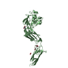
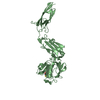
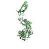
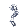
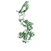
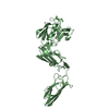
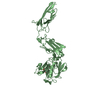
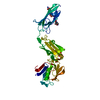
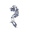
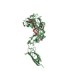
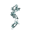
- Structure visualization
| Structure viewer | Molecule: MolmilJmol/JSmol |
|---|
- Downloads & links
-Download
| PDBx/mmCIF format | 5dvp.cif.gz | 150.9 KB | Display | PDBx/mmCIF format |
|---|---|---|---|---|
| PDB format | pdb5dvp.ent.gz | 115.3 KB | Display | PDB format |
| PDBx/mmJSON format | 5dvp.json.gz | Tree view | PDBx/mmJSON format | |
| Others |  Other downloads Other downloads |
-Validation report
| Arichive directory | https://data.pdbj.org/pub/pdb/validation_reports/dv/5dvpftp://data.pdbj.org/pub/pdb/validation_reports/dv/5dvp | HTTPS FTP |
|---|
-Related structure data
| Related structure data |  5du7C  5dujC  5dzjC  5dzpC  5e1gC  5e1iC  5e51C  5e5lC  5k69C  3vynS  5dvq C: citing same article ( S: Starting model for refinement |
|---|---|
| Similar structure data |


-Links
 PDBj
PDBj


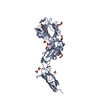
- Assembly
| Deposited unit | 
| ||||||||
|---|---|---|---|---|---|---|---|---|---|
| 1 | 
| ||||||||
| 2 | 
| ||||||||
| Unit cell |
|
-Components
| #1: Protein | Mass: 37494.609 Da / Num. of mol.: 2 / Fragment: residues 58-407 Source method: isolated from a genetically manipulated source Source: (gene. exp.) Mycobacterium tuberculosis (bacteria) / Strain: ATCC 25618 / H37Rv / Gene: ldtB, lppS, Rv2518c, RVBD_2518c, P425_02624 / Plasmid: pET28a / Production host: References: UniProt: I6Y9J2, Transferases; Acyltransferases; Aminoacyltransferases #2: Chemical | 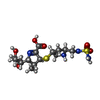  Mass: 422.520 Da / Num. of mol.: 2 / Source method: obtained synthetically / Formula: C15H26N4O6S2 Mass: 422.520 Da / Num. of mol.: 2 / Source method: obtained synthetically / Formula: C15H26N4O6S2#3: Chemical | ChemComp-SO4 / |   Mass: 96.063 Da / Num. of mol.: 1 / Source method: obtained synthetically / Formula: SO4 Mass: 96.063 Da / Num. of mol.: 1 / Source method: obtained synthetically / Formula: SO4#4: Chemical | 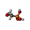  Mass: 124.032 Da / Num. of mol.: 2 / Source method: obtained synthetically / Formula: C2H5O4P Mass: 124.032 Da / Num. of mol.: 2 / Source method: obtained synthetically / Formula: C2H5O4P#5: Water | ChemComp-HOH / |  Mass: 18.015 Da / Num. of mol.: 232 / Source method: isolated from a natural source / Formula: H2O Mass: 18.015 Da / Num. of mol.: 232 / Source method: isolated from a natural source / Formula: H2OHas protein modification | Y | |
|---|
-Experimental details
-Experiment
| Experiment | Method: X-RAY DIFFRACTION / Number of used crystals: 1 |
|---|
- Sample preparation
| Crystal | Density Matthews: 2.84 Å3/Da / Density % sol: 56.64 % |
|---|---|
| Crystal grow | Temperature: 291 K / Method: vapor diffusion, hanging drop / Details: 20% 5000MME, 200mM Ammonium sulfate |
-Data collection
| Diffraction | Mean temperature: 100 K |
|---|---|
| Diffraction source | Source: ROTATING ANODE / Type: RIGAKU / Wavelength: 1.5 Å |
| Detector | Type: RIGAKU SATURN 944+ / Detector: CCD / Date: Oct 7, 2014 |
| Radiation | Protocol: SINGLE WAVELENGTH / Monochromatic (M) / Laue (L): M / Scattering type: x-ray |
| Radiation wavelength | Wavelength: 1.5 Å / Relative weight: 1 |
| Reflection | Resolution: 2.18→50 Å / Num. obs: 43922 / % possible obs: 99.7 % / Redundancy: 3.7 % / Rmerge(I) obs: 0.061 / Rsym value: 0.05 / Net I/σ(I): 15.84 |
| Reflection shell | Resolution: 2.18→2.22 Å / Redundancy: 2.7 % / Rmerge(I) obs: 0.218 / Mean I/σ(I) obs: 2.69 / % possible all: 95.5 |
- Processing
| Software |
| |||||||||||||||||||||||||||||||||||||||||||||||||||||||||||||||||||||||||||||||||||||||||||||||||||||||||
|---|---|---|---|---|---|---|---|---|---|---|---|---|---|---|---|---|---|---|---|---|---|---|---|---|---|---|---|---|---|---|---|---|---|---|---|---|---|---|---|---|---|---|---|---|---|---|---|---|---|---|---|---|---|---|---|---|---|---|---|---|---|---|---|---|---|---|---|---|---|---|---|---|---|---|---|---|---|---|---|---|---|---|---|---|---|---|---|---|---|---|---|---|---|---|---|---|---|---|---|---|---|---|---|---|---|---|
| Refinement | Method to determine structure: MOLECULAR REPLACEMENT Starting model: 3VYN Resolution: 2.18→41.324 Å / SU ML: 0.24 / Cross valid method: FREE R-VALUE / σ(F): 1.39 / Phase error: 21.92 / Stereochemistry target values: ML
| |||||||||||||||||||||||||||||||||||||||||||||||||||||||||||||||||||||||||||||||||||||||||||||||||||||||||
| Solvent computation | Shrinkage radii: 0.9 Å / VDW probe radii: 1.11 Å / Solvent model: FLAT BULK SOLVENT MODEL | |||||||||||||||||||||||||||||||||||||||||||||||||||||||||||||||||||||||||||||||||||||||||||||||||||||||||
| Displacement parameters | Biso max: 64.49 Å2 / Biso mean: 24.7835 Å2 / Biso min: 10.67 Å2 | |||||||||||||||||||||||||||||||||||||||||||||||||||||||||||||||||||||||||||||||||||||||||||||||||||||||||
| Refinement step | Cycle: final / Resolution: 2.18→41.324 Å
| |||||||||||||||||||||||||||||||||||||||||||||||||||||||||||||||||||||||||||||||||||||||||||||||||||||||||
| Refine LS restraints |
| |||||||||||||||||||||||||||||||||||||||||||||||||||||||||||||||||||||||||||||||||||||||||||||||||||||||||
| LS refinement shell | Refine-ID: X-RAY DIFFRACTION / Total num. of bins used: 14
|
