 Movie
Movie Controller

[English] 日本語
 Yorodumi
- PDB-5uwv: Crystal structure of Mycobacterium abscessus L,D-transpeptidase 2 -
+ Open data

- Basic information
| Entry | Database: PDB / ID: 5uwv | ||||||
|---|---|---|---|---|---|---|---|
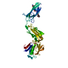
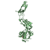
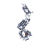
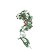
| Title | Crystal structure of Mycobacterium abscessus L,D-transpeptidase 2 | ||||||
 Components Components | L,D-TRANSPEPTIDASE 2 | ||||||
 Keywords Keywords | TRANSFERASE / L / D-transpeptidase / Peptidoglycan synthesis enzyme / cell wall enzyme / LdtMab2 / Mycobacterium abscessus | ||||||
| Function / homology |  Function and homology information Function and homology informationacyltransferase activity / peptidoglycan biosynthetic process / cell wall organization / regulation of cell shape Similarity search - Function | ||||||
| Biological species |  Mycobacterium abscessus (bacteria) Mycobacterium abscessus (bacteria) | ||||||
| Method |  X-RAY DIFFRACTION / SYNCHROTRON / MOLECULAR REPLACEMENT / Resolution: 2.98 Å X-RAY DIFFRACTION / SYNCHROTRON / MOLECULAR REPLACEMENT / Resolution: 2.98 Å | ||||||
 Authors Authors | Kumar, P. / Ginell, S.L. / Lamichhane, G. | ||||||
| Funding support |  United States, 1items United States, 1items
| ||||||
 Citation Citation | Journal: Antimicrob. Agents Chemother. / Year: 2017 Title: Mycobacterium abscessus l,d-Transpeptidases Are Susceptible to Inactivation by Carbapenems and Cephalosporins but Not Penicillins. Authors: Kumar, P. / Chauhan, V. / Silva, J.R.A. / Lameira, J. / d'Andrea, F.B. / Li, S.G. / Ginell, S.L. / Freundlich, J.S. / Alves, C.N. / Bailey, S. / Cohen, K.A. / Lamichhane, G. | ||||||
| History |
|
- Structure visualization
| Structure viewer | Molecule: MolmilJmol/JSmol |
|---|
- Downloads & links
-Download
| PDBx/mmCIF format | 5uwv.cif.gz | 378.6 KB | Display | PDBx/mmCIF format |
|---|---|---|---|---|
| PDB format | pdb5uwv.ent.gz | 308.2 KB | Display | PDB format |
| PDBx/mmJSON format | 5uwv.json.gz | Tree view | PDBx/mmJSON format | |
| Others |  Other downloads Other downloads |
-Validation report
| Arichive directory | https://data.pdbj.org/pub/pdb/validation_reports/uw/5uwvftp://data.pdbj.org/pub/pdb/validation_reports/uw/5uwv | HTTPS FTP |
|---|
-Related structure data
| Related structure data |  5du7S S: Starting model for refinement |
|---|---|
| Similar structure data |


-Links
 PDBj
PDBj


- Assembly
| Deposited unit | 
| ||||||||
|---|---|---|---|---|---|---|---|---|---|
| 1 | 
| ||||||||
| 2 | 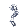
| ||||||||
| 3 | 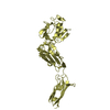
| ||||||||
| 4 | 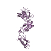
| ||||||||
| 5 | 
| ||||||||
| 6 | 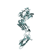
| ||||||||
| Unit cell |
|
-Components
| #1: Protein | Mass: 39522.059 Da / Num. of mol.: 6 / Fragment: UNP residues 42-406 Source method: isolated from a genetically manipulated source Source: (gene. exp.) Mycobacterium abscessus (strain ATCC 19977 / DSM 44196 / CIP 104536 / JCM 13569 / NCTC 13031 / TMC 1543) (bacteria)Strain: ATCC 19977 / DSM 44196 / CIP 104536 / JCM 13569 / NCTC 13031 / TMC 1543 Gene: MAB_1530 / Plasmid: pET28a / Production host: #2: Water | ChemComp-HOH / |  Mass: 18.015 Da / Num. of mol.: 60 / Source method: isolated from a natural source / Formula: H2O Mass: 18.015 Da / Num. of mol.: 60 / Source method: isolated from a natural source / Formula: H2O |
|---|
-Experimental details
-Experiment
| Experiment | Method: X-RAY DIFFRACTION / Number of used crystals: 1 |
|---|
- Sample preparation
| Crystal | Density Matthews: 3.53 Å3/Da / Density % sol: 65.12 % |
|---|---|
| Crystal grow | Temperature: 291 K / Method: vapor diffusion, hanging drop Details: 13% PEG 8000, 110 mM sodium citrate tribasic dihydrate PH range: pH 5.5 |
-Data collection
| Diffraction | Mean temperature: 100 K |
|---|---|
| Diffraction source | Source: SYNCHROTRON / Site: APS / Beamline: 19-ID / Wavelength: 0.97918 Å |
| Detector | Type: DECTRIS PILATUS3 6M / Detector: PIXEL / Date: Mar 20, 2015 / Details: mirrors |
| Radiation | Monochromator: Si(111) / Protocol: SINGLE WAVELENGTH / Monochromatic (M) / Laue (L): M / Scattering type: x-ray |
| Radiation wavelength | Wavelength: 0.97918 Å / Relative weight: 1 |
| Reflection | Resolution: 2.98→135.53 Å / Num. obs: 65870 / % possible obs: 99.4 % / Redundancy: 4.6 % / Biso Wilson estimate: 56.54 Å2 / CC1/2: 0.96 / Rmerge(I) obs: 0.06 / Net I/σ(I): 21.02 |
| Reflection shell | Resolution: 2.98→3.087 Å / Redundancy: 4.8 % / Rmerge(I) obs: 0.57 / Mean I/σ(I) obs: 2.52 / Num. unique obs: 5838 / CC1/2: 0.78 / % possible all: 99.9 |
- Processing
| Software |
| ||||||||||||||||||||||||||||||||||||||||||||||||||||||||||||
|---|---|---|---|---|---|---|---|---|---|---|---|---|---|---|---|---|---|---|---|---|---|---|---|---|---|---|---|---|---|---|---|---|---|---|---|---|---|---|---|---|---|---|---|---|---|---|---|---|---|---|---|---|---|---|---|---|---|---|---|---|---|
| Refinement | Method to determine structure: MOLECULAR REPLACEMENT Starting model: 5DU7 Resolution: 2.98→135.53 Å / Cor.coef. Fo:Fc: 0.928 / Cor.coef. Fo:Fc free: 0.874 / SU B: 18.041 / SU ML: 0.317 / Cross valid method: THROUGHOUT / σ(F): 0 / ESU R: 0.565 / ESU R Free: 0.409 Details: HYDROGENS HAVE BEEN ADDED IN THE RIDING POSITIONS U VALUES : REFINED INDIVIDUALLY
| ||||||||||||||||||||||||||||||||||||||||||||||||||||||||||||
| Solvent computation | Ion probe radii: 0.8 Å / Shrinkage radii: 0.8 Å / VDW probe radii: 1.2 Å | ||||||||||||||||||||||||||||||||||||||||||||||||||||||||||||
| Displacement parameters | Biso max: 138.68 Å2 / Biso mean: 58.908 Å2 / Biso min: 20 Å2
| ||||||||||||||||||||||||||||||||||||||||||||||||||||||||||||
| Refinement step | Cycle: final / Resolution: 2.98→135.53 Å
| ||||||||||||||||||||||||||||||||||||||||||||||||||||||||||||
| Refine LS restraints |
| ||||||||||||||||||||||||||||||||||||||||||||||||||||||||||||
| LS refinement shell | Resolution: 2.981→3.058 Å / Rfactor Rfree error: 0 / Total num. of bins used: 20
|
