[English] 日本語
Yorodumi
- PDB-4qqr: Structural insight into nucleotide rhamnose synthase/epimerase-re... -

+
Open data


ID or keywords:

Loading...

-
Basic information

Entry
Database: PDB / ID: 4qqr
TitleStructural insight into nucleotide rhamnose synthase/epimerase-reductase from Arabidopsis thaliana
Components3,5-epimerase/4-reductase
KeywordsOXIDOREDUCTASE / BETA BARREL / Rossmann Fold / epimerase-reductase
Function / homology
Function and homology information


UDP-rhamnose biosynthetic process / UDP-4-keto-6-deoxy-glucose-3,5-epimerase activity / UDP-4-keto-rhamnose-4-keto-reductase activity / dTDP-4-dehydrorhamnose reductase / dTDP-4-dehydrorhamnose reductase activity / dTDP-4-dehydrorhamnose 3,5-epimerase / dTDP-4-dehydrorhamnose 3,5-epimerase activity / dTDP-rhamnose biosynthetic process / plasmodesma / oxidoreductase activity, acting on the CH-OH group of donors, NAD or NADP as acceptor ...UDP-rhamnose biosynthetic process / UDP-4-keto-6-deoxy-glucose-3,5-epimerase activity / UDP-4-keto-rhamnose-4-keto-reductase activity / dTDP-4-dehydrorhamnose reductase / dTDP-4-dehydrorhamnose reductase activity / dTDP-4-dehydrorhamnose 3,5-epimerase / dTDP-4-dehydrorhamnose 3,5-epimerase activity / dTDP-rhamnose biosynthetic process / plasmodesma / oxidoreductase activity, acting on the CH-OH group of donors, NAD or NADP as acceptor / cell wall organization / oxidoreductase activity / plasma membrane / cytosol
Similarity search - Function
RmlD-like substrate binding domain / RmlD substrate binding domain / NAD(P)-binding Rossmann-like Domain / NAD(P)-binding domain superfamily / Rossmann fold / 3-Layer(aba) Sandwich / Alpha Beta
Similarity search - Domain/homology
Bifunctional dTDP-4-dehydrorhamnose 3,5-epimerase/dTDP-4-dehydrorhamnose reductase
Similarity search - Component
Biological speciesArabidopsis thaliana (thale cress)
MethodX-RAY DIFFRACTION / SYNCHROTRON / SAD / Resolution: 2.7 Å
AuthorsHan, X. / Liu, X.
Citation
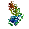
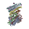
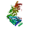
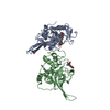
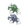
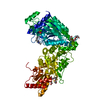
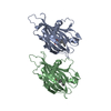
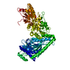
Journal: To be Published
Title: Structural insight into nucleotide rhamnose synthase/epimerase-reductase from Arabidopsis thaliana
Authors: Han, X. / Liu, X.
History
DepositionJun 28, 2014Deposition site: RCSB / Processing site: PDBJ
Revision 1.0Jul 1, 2015Provider: repository / Type: Initial release
Revision 1.1Mar 20, 2024Group: Data collection / Database references / Derived calculations
Category: chem_comp_atom / chem_comp_bond ...chem_comp_atom / chem_comp_bond / database_2 / pdbx_struct_conn_angle / struct_conn / struct_site
Item: _database_2.pdbx_DOI / _database_2.pdbx_database_accession ..._database_2.pdbx_DOI / _database_2.pdbx_database_accession / _pdbx_struct_conn_angle.ptnr1_auth_asym_id / _pdbx_struct_conn_angle.ptnr1_auth_comp_id / _pdbx_struct_conn_angle.ptnr1_auth_seq_id / _pdbx_struct_conn_angle.ptnr1_label_asym_id / _pdbx_struct_conn_angle.ptnr1_label_atom_id / _pdbx_struct_conn_angle.ptnr1_label_comp_id / _pdbx_struct_conn_angle.ptnr1_label_seq_id / _pdbx_struct_conn_angle.ptnr2_auth_asym_id / _pdbx_struct_conn_angle.ptnr2_auth_seq_id / _pdbx_struct_conn_angle.ptnr2_label_asym_id / _pdbx_struct_conn_angle.ptnr3_auth_asym_id / _pdbx_struct_conn_angle.ptnr3_auth_comp_id / _pdbx_struct_conn_angle.ptnr3_auth_seq_id / _pdbx_struct_conn_angle.ptnr3_label_asym_id / _pdbx_struct_conn_angle.ptnr3_label_atom_id / _pdbx_struct_conn_angle.ptnr3_label_comp_id / _pdbx_struct_conn_angle.ptnr3_label_seq_id / _pdbx_struct_conn_angle.value / _struct_conn.pdbx_dist_value / _struct_conn.ptnr1_auth_asym_id / _struct_conn.ptnr1_auth_comp_id / _struct_conn.ptnr1_auth_seq_id / _struct_conn.ptnr1_label_asym_id / _struct_conn.ptnr1_label_atom_id / _struct_conn.ptnr1_label_comp_id / _struct_conn.ptnr1_label_seq_id / _struct_conn.ptnr2_auth_asym_id / _struct_conn.ptnr2_auth_comp_id / _struct_conn.ptnr2_auth_seq_id / _struct_conn.ptnr2_label_asym_id / _struct_conn.ptnr2_label_atom_id / _struct_conn.ptnr2_label_comp_id / _struct_conn.ptnr2_label_seq_id / _struct_site.pdbx_auth_asym_id / _struct_site.pdbx_auth_comp_id / _struct_site.pdbx_auth_seq_id

-
Structure visualization

Structure viewerMolecule:
MolmilJmol/JSmol

Downloads & links

-
Assembly

Deposited unit
A: 3,5-epimerase/4-reductase
B: 3,5-epimerase/4-reductase
hetero molecules


Theoretical massNumber of molelcules
Total (without water)67,89312
Polymers67,2692
Non-polymers62510
Water1,22568
1


  • Idetical with deposited unit
  • defined by author&software
TypeNameSymmetry operationNumber
identity operation1_555x,y,z1
Buried area3280 Å2
ΔGint-242 kcal/mol
Surface area23050 Å2
MethodPISA
Unit cell
Length a, b, c (Å)56.589, 61.943, 176.774
Angle α, β, γ (deg.)90.00, 90.00, 90.00
Int Tables number19
Space group name H-MP212121

-
Components

#1: Protein 3,5-epimerase/4-reductase / F16P17.17 protein / Putative uncharacterized protein At1g63000 / UDP-4-keto-6-deoxy-d-glucose-3 / 5- ...F16P17.17 protein / Putative uncharacterized protein At1g63000 / UDP-4-keto-6-deoxy-d-glucose-3 / 5-epimerase-4-reductase 1


Mass: 33634.258 Da / Num. of mol.: 2
Source method: isolated from a genetically manipulated source
Source: (gene. exp.) Arabidopsis thaliana (thale cress) / Gene: At1g63000, ER, F16P17.17, NRS, NRS/ER / Production host: Escherichia coli (E. coli) / References: UniProt: Q9LQ04
#2: Chemical
ChemComp-ZN / ZINC ION


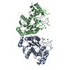
Mass: 65.409 Da / Num. of mol.: 7 / Source method: obtained synthetically / Formula: Zn
#3: Chemical ChemComp-CL / CHLORIDE ION / Chloride


Mass: 35.453 Da / Num. of mol.: 2 / Source method: obtained synthetically / Formula: Cl
#4: Chemical ChemComp-SO4 / SULFATE ION / Sulfate


Mass: 96.063 Da / Num. of mol.: 1 / Source method: obtained synthetically / Formula: SO4
#5: Water ChemComp-HOH / water / Water


Mass: 18.015 Da / Num. of mol.: 68 / Source method: isolated from a natural source / Formula: H2O

-
Experimental details

-
Experiment

ExperimentMethod: X-RAY DIFFRACTION / Number of used crystals: 4

-
Sample preparation

CrystalDensity Matthews: 2.3 Å3/Da / Density % sol: 46.59 %
Description: THE ENTRY CONTAINS FRIEDEL PAIRS IN F_PLUS/MINUS COLUMNS
Crystal growTemperature: 277 K / Method: vapor diffusion, sitting drop / pH: 6.5
Details: 1.8M ammonium sulfate, 0.1mM ZnCl2, 1mM DTT and 0.1M Hepes pH 6.5. , VAPOR DIFFUSION, SITTING DROP, temperature 277K

-
Data collection

DiffractionMean temperature: 298 K
Diffraction sourceSource: SYNCHROTRON / Site: SPring-8 / Beamline: BL12B2 / Wavelength: 1.2826 Å
DetectorType: ADSC QUANTUM 1 / Detector: CCD / Date: Nov 15, 2012
RadiationMonochromator: GRAPHITE / Protocol: SINGLE WAVELENGTH / Monochromatic (M) / Laue (L): M / Scattering type: x-ray
Radiation wavelengthWavelength: 1.2826 Å / Relative weight: 1
ReflectionResolution: 2.7→50 Å / Num. obs: 34737 / % possible obs: 95 % / Observed criterion σ(F): 2 / Observed criterion σ(I): 2
Reflection shell
Resolution (Å)Diffraction-ID% possible all
2.7-4.131100
4.13-7.051100
7.05-501100

-
Processing

SoftwareName: PHENIX / Version: (phenix.refine: 1.7.3_928) / Classification: refinement
RefinementMethod to determine structure: SAD / Resolution: 2.7→41.779 Å / SU ML: 0.38 / σ(F): 1.34 / Phase error: 27.68 / Stereochemistry target values: ML
Details: SF FILE CONTAINS FRIEDEL PAIRS UNDER I/F_MINUS AND I/F_PLUS COLUMNS.
RfactorNum. reflection% reflectionSelection details
Rfree0.2652 3485 10.03 %RANDOM
Rwork0.1985 ---
all0.247 ---
obs0.2051 34735 94.42 %-
Solvent computationShrinkage radii: 0.73 Å / VDW probe radii: 1 Å / Solvent model: FLAT BULK SOLVENT MODEL / Bsol: 43.022 Å2 / ksol: 0.352 e/Å3
Displacement parameters
Baniso -1Baniso -2Baniso -3
1-1.3851 Å20 Å2-0 Å2
2--6.5156 Å2-0 Å2
3----7.9008 Å2
Refinement stepCycle: LAST / Resolution: 2.7→41.779 Å
ProteinNucleic acidLigandSolventTotal
Num. atoms4270 0 14 68 4352
Refine LS restraints
Refine-IDTypeDev idealNumber
X-RAY DIFFRACTIONf_bond_d0.0094358
X-RAY DIFFRACTIONf_angle_d1.1795899
X-RAY DIFFRACTIONf_dihedral_angle_d15.961599
X-RAY DIFFRACTIONf_chiral_restr0.078668
X-RAY DIFFRACTIONf_plane_restr0.007748
LS refinement shell

Refine-ID: X-RAY DIFFRACTION / Total num. of bins used: 25

Resolution (Å)Rfactor RfreeNum. reflection RfreeRfactor RworkNum. reflection Rwork% reflection obs (%)
2.603-2.63860.40121310.2668116889
2.6386-2.67630.36531370.2711122092
2.6763-2.71630.34271470.2531128395
2.7163-2.75870.3511470.2645125998
2.7587-2.80390.35311540.25971347100
2.8039-2.85230.30411420.25451309100
2.8523-2.90410.35671500.24161316100
2.9041-2.960.34041510.24751340100
2.96-3.02040.33331430.23961308100
3.0204-3.0860.33191540.24681360100
3.086-3.15780.32621420.21541294100
3.1578-3.23670.32011470.23091336100
3.2367-3.32420.24951500.19451332100
3.3242-3.4220.31671470.20631308100
3.422-3.53240.31031470.20481329100
3.5324-3.65860.3955950.200286395
3.6586-3.80490.2298770.23172994
3.8049-3.9780.25141440.206124195
3.978-4.18750.25881400.1681129298
4.1875-4.44960.19281420.1489130997
4.4496-4.79270.16981440.1351127097
4.7927-5.27420.20091430.1569130599
5.2742-6.03540.24061420.176131899
6.0354-7.59650.23931440.1903130198
7.5965-41.78470.21511250.2068111584

+
About Yorodumi

-
News

-
Feb 9, 2022. New format data for meta-information of EMDB entries

New format data for meta-information of EMDB entries

  • Version 3 of the EMDB header file is now the official format.
  • The previous official version 1.9 will be removed from the archive.

Related info.:EMDB header

External links:wwPDB to switch to version 3 of the EMDB data model

-
Aug 12, 2020. Covid-19 info

Covid-19 info

URL: https://pdbj.org/emnavi/covid19.php

New page: Covid-19 featured information page in EM Navigator.

Related info.:Covid-19 info / Mar 5, 2020. Novel coronavirus structure data

+
Mar 5, 2020. Novel coronavirus structure data

Novel coronavirus structure data

Related info.:Yorodumi Speices / Aug 12, 2020. Covid-19 info

External links:COVID-19 featured content - PDBj / Molecule of the Month (242):Coronavirus Proteases

+
Jan 31, 2019. EMDB accession codes are about to change! (news from PDBe EMDB page)

EMDB accession codes are about to change! (news from PDBe EMDB page)

  • The allocation of 4 digits for EMDB accession codes will soon come to an end. Whilst these codes will remain in use, new EMDB accession codes will include an additional digit and will expand incrementally as the available range of codes is exhausted. The current 4-digit format prefixed with “EMD-” (i.e. EMD-XXXX) will advance to a 5-digit format (i.e. EMD-XXXXX), and so on. It is currently estimated that the 4-digit codes will be depleted around Spring 2019, at which point the 5-digit format will come into force.
  • The EM Navigator/Yorodumi systems omit the EMD- prefix.

Related info.:Q: What is EMD? / ID/Accession-code notation in Yorodumi/EM Navigator

External links:EMDB Accession Codes are Changing Soon! / Contact to PDBj

+
Jul 12, 2017. Major update of PDB

Major update of PDB

  • wwPDB released updated PDB data conforming to the new PDBx/mmCIF dictionary.
  • This is a major update changing the version number from 4 to 5, and with Remediation, in which all the entries are updated.
  • In this update, many items about electron microscopy experimental information are reorganized (e.g. em_software).
  • Now, EM Navigator and Yorodumi are based on the updated data.

External links:wwPDB Remediation / Enriched Model Files Conforming to OneDep Data Standards Now Available in the PDB FTP Archive

-
Yorodumi

Thousand views of thousand structures

  • Yorodumi is a browser for structure data from EMDB, PDB, SASBDB, etc.
  • This page is also the successor to EM Navigator detail page, and also detail information page/front-end page for Omokage search.
  • The word "yorodu" (or yorozu) is an old Japanese word meaning "ten thousand". "mi" (miru) is to see.

Related info.:EMDB / PDB / SASBDB / Comparison of 3 databanks / Yorodumi Search / Aug 31, 2016. New EM Navigator & Yorodumi / Yorodumi Papers / Jmol/JSmol / Function and homology information / Changes in new EM Navigator and Yorodumi

Read more