 Movie
Movie Controller

[English] 日本語
 Yorodumi
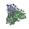
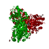
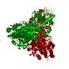
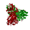
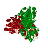
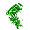
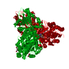
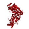
- PDB-5hyv: Crystal Structure of Transketolase with ThDP from Pichia Stipitis -
+ Open data

- Basic information
| Entry | Database: PDB / ID: 5hyv | ||||||
|---|---|---|---|---|---|---|---|
| Title | Crystal Structure of Transketolase with ThDP from Pichia Stipitis | ||||||
 Components Components | Transketolase | ||||||
 Keywords Keywords | TRANSFERASE / transketolase | ||||||
| Function / homology |  Function and homology information Function and homology informationtransketolase / transketolase activity / pentose-phosphate shunt / metal ion binding / nucleus / cytosol Similarity search - Function | ||||||
| Biological species |  Scheffersomyces stipitis CBS 6054 (fungus) Scheffersomyces stipitis CBS 6054 (fungus) | ||||||
| Method |  X-RAY DIFFRACTION / SYNCHROTRON / MOLECULAR REPLACEMENT / Resolution: 1.031 Å X-RAY DIFFRACTION / SYNCHROTRON / MOLECULAR REPLACEMENT / Resolution: 1.031 Å | ||||||
 Authors Authors | Li, T.L. / Hsu, L.J. / Hsu, N.S. | ||||||
 Citation Citation | Journal: Protein Eng. Des. Sel. / Year: 2016 Title: Structural and biochemical interrogation on transketolase from Pichia stipitis for new functionality Authors: Hsu, L.J. / Hsu, N.S. / Wang, Y.L. / Wu, C.J. / Li, T.L. | ||||||
| History |
|
- Structure visualization
| Structure viewer | Molecule: MolmilJmol/JSmol |
|---|
- Downloads & links
-Download
| PDBx/mmCIF format | 5hyv.cif.gz | 425.8 KB | Display | PDBx/mmCIF format |
|---|---|---|---|---|
| PDB format | pdb5hyv.ent.gz | 346.7 KB | Display | PDB format |
| PDBx/mmJSON format | 5hyv.json.gz | Tree view | PDBx/mmJSON format | |
| Others |  Other downloads Other downloads |
-Validation report
| Arichive directory | https://data.pdbj.org/pub/pdb/validation_reports/hy/5hyvftp://data.pdbj.org/pub/pdb/validation_reports/hy/5hyv | HTTPS FTP |
|---|
-Related structure data
| Related structure data |  5hgxC  5hjeC  5i4iC  5i51C  5i5eC  5i5gC 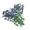 1gpuS C: citing same article ( S: Starting model for refinement |
|---|---|
| Similar structure data |


-Links
 PDBj
PDBj


- Assembly
| Deposited unit | 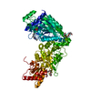
| ||||||||||||||||||||||||
|---|---|---|---|---|---|---|---|---|---|---|---|---|---|---|---|---|---|---|---|---|---|---|---|---|---|
| 1 | 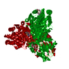
| ||||||||||||||||||||||||
| Unit cell |
| ||||||||||||||||||||||||
| Components on special symmetry positions |
|
-Components
| #1: Protein | Mass: 75054.453 Da / Num. of mol.: 1 Source method: isolated from a genetically manipulated source Source: (gene. exp.) Scheffersomyces stipitis CBS 6054 (fungus)Strain: CBS 6054 / Gene: TKT, TKT1, PICST_67105 / Production host:  | ||
|---|---|---|---|
| #2: Chemical | ChemComp-TPP /   Mass: 425.314 Da / Num. of mol.: 1 / Source method: obtained synthetically / Formula: C12H19N4O7P2S Mass: 425.314 Da / Num. of mol.: 1 / Source method: obtained synthetically / Formula: C12H19N4O7P2S | ||
| #3: Chemical | ChemComp-CA /   Mass: 40.078 Da / Num. of mol.: 1 / Source method: obtained synthetically / Formula: Ca Mass: 40.078 Da / Num. of mol.: 1 / Source method: obtained synthetically / Formula: Ca | ||
| #4: Chemical |   Mass: 238.278 Da / Num. of mol.: 2 / Source method: obtained synthetically / Formula: C10H22O6 / Comment: precipitant*YM Mass: 238.278 Da / Num. of mol.: 2 / Source method: obtained synthetically / Formula: C10H22O6 / Comment: precipitant*YM#5: Water | ChemComp-HOH / |  Mass: 18.015 Da / Num. of mol.: 899 / Source method: isolated from a natural source / Formula: H2O Mass: 18.015 Da / Num. of mol.: 899 / Source method: isolated from a natural source / Formula: H2O |
-Experimental details
-Experiment
| Experiment | Method: X-RAY DIFFRACTION / Number of used crystals: 1 |
|---|
- Sample preparation
| Crystal | Density Matthews: 3.15 Å3/Da / Density % sol: 60.96 % |
|---|---|
| Crystal grow | Temperature: 293 K / Method: vapor diffusion, hanging drop / Details: MES, NaCl, PEG400 |
-Data collection
| Diffraction | Mean temperature: 100 K | ||||||||||||||||||||||||||||||||||||||||||||||||||||||||||||||||||
|---|---|---|---|---|---|---|---|---|---|---|---|---|---|---|---|---|---|---|---|---|---|---|---|---|---|---|---|---|---|---|---|---|---|---|---|---|---|---|---|---|---|---|---|---|---|---|---|---|---|---|---|---|---|---|---|---|---|---|---|---|---|---|---|---|---|---|---|
| Diffraction source | Source: SYNCHROTRON / Site: NSRRC  / Beamline: BL13C1 / Wavelength: 0.974 Å / Beamline: BL13C1 / Wavelength: 0.974 Å | ||||||||||||||||||||||||||||||||||||||||||||||||||||||||||||||||||
| Detector | Type: ADSC QUANTUM 315 / Detector: CCD / Date: Sep 22, 2012 | ||||||||||||||||||||||||||||||||||||||||||||||||||||||||||||||||||
| Radiation | Protocol: SINGLE WAVELENGTH / Monochromatic (M) / Laue (L): M / Scattering type: x-ray | ||||||||||||||||||||||||||||||||||||||||||||||||||||||||||||||||||
| Radiation wavelength | Wavelength: 0.974 Å / Relative weight: 1 | ||||||||||||||||||||||||||||||||||||||||||||||||||||||||||||||||||
| Reflection | Resolution: 1.03→30 Å / Num. obs: 443954 / % possible obs: 99.3 % / Redundancy: 7 % / Biso Wilson estimate: 10.42 Å2 / Rmerge(I) obs: 0.075 / Χ2: 1.147 / Net I/av σ(I): 18.787 / Net I/σ(I): 16 / Num. measured all: 3096480 | ||||||||||||||||||||||||||||||||||||||||||||||||||||||||||||||||||
| Reflection shell | Diffraction-ID: 1 / Rejects: _
|
- Processing
| Software |
| |||||||||||||||||||||||||||||||||||||||||||||||||||||||||||||||||||||||||||||||||||||||||||||||||||||||||||||||||||||||||||||||||||||||||||||||||||||||||||||||||||||||||||||||||||||||||||||||||||||||||||||||||||||||||
|---|---|---|---|---|---|---|---|---|---|---|---|---|---|---|---|---|---|---|---|---|---|---|---|---|---|---|---|---|---|---|---|---|---|---|---|---|---|---|---|---|---|---|---|---|---|---|---|---|---|---|---|---|---|---|---|---|---|---|---|---|---|---|---|---|---|---|---|---|---|---|---|---|---|---|---|---|---|---|---|---|---|---|---|---|---|---|---|---|---|---|---|---|---|---|---|---|---|---|---|---|---|---|---|---|---|---|---|---|---|---|---|---|---|---|---|---|---|---|---|---|---|---|---|---|---|---|---|---|---|---|---|---|---|---|---|---|---|---|---|---|---|---|---|---|---|---|---|---|---|---|---|---|---|---|---|---|---|---|---|---|---|---|---|---|---|---|---|---|---|---|---|---|---|---|---|---|---|---|---|---|---|---|---|---|---|---|---|---|---|---|---|---|---|---|---|---|---|---|---|---|---|---|---|---|---|---|---|---|---|---|---|---|---|---|---|---|---|---|
| Refinement | Method to determine structure: MOLECULAR REPLACEMENT Starting model: 1GPU Resolution: 1.031→27.603 Å / SU ML: 0.09 / Cross valid method: FREE R-VALUE / σ(F): 1.34 / Phase error: 9.29
| |||||||||||||||||||||||||||||||||||||||||||||||||||||||||||||||||||||||||||||||||||||||||||||||||||||||||||||||||||||||||||||||||||||||||||||||||||||||||||||||||||||||||||||||||||||||||||||||||||||||||||||||||||||||||
| Solvent computation | Shrinkage radii: 0.9 Å / VDW probe radii: 1.11 Å | |||||||||||||||||||||||||||||||||||||||||||||||||||||||||||||||||||||||||||||||||||||||||||||||||||||||||||||||||||||||||||||||||||||||||||||||||||||||||||||||||||||||||||||||||||||||||||||||||||||||||||||||||||||||||
| Displacement parameters | Biso max: 66.04 Å2 / Biso mean: 15.3387 Å2 / Biso min: 7.65 Å2 | |||||||||||||||||||||||||||||||||||||||||||||||||||||||||||||||||||||||||||||||||||||||||||||||||||||||||||||||||||||||||||||||||||||||||||||||||||||||||||||||||||||||||||||||||||||||||||||||||||||||||||||||||||||||||
| Refinement step | Cycle: final / Resolution: 1.031→27.603 Å
| |||||||||||||||||||||||||||||||||||||||||||||||||||||||||||||||||||||||||||||||||||||||||||||||||||||||||||||||||||||||||||||||||||||||||||||||||||||||||||||||||||||||||||||||||||||||||||||||||||||||||||||||||||||||||
| Refine LS restraints |
| |||||||||||||||||||||||||||||||||||||||||||||||||||||||||||||||||||||||||||||||||||||||||||||||||||||||||||||||||||||||||||||||||||||||||||||||||||||||||||||||||||||||||||||||||||||||||||||||||||||||||||||||||||||||||
| LS refinement shell | Refine-ID: X-RAY DIFFRACTION / Total num. of bins used: 30
|
