 Movie
Movie Controller


+ Open data

- Basic information
| Entry | Database: PDB / ID: 4qc7 | ||||||
|---|---|---|---|---|---|---|---|
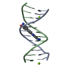
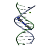
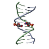
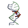
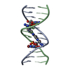
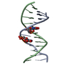
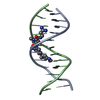
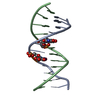
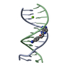
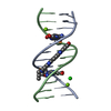
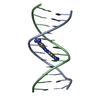
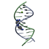
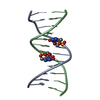
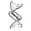
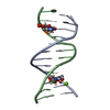
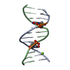
| Title | Dodecamer structure of 5-formylcytosine containing DNA | ||||||
 Components Components | short DNA strands | ||||||
 Keywords Keywords | DNA / double helix / B-form DNA / 5-formylcytosine | ||||||
| Function / homology | DNA / DNA (> 10) Function and homology information Function and homology information | ||||||
| Method |  X-RAY DIFFRACTION / SYNCHROTRON / MOLECULAR REPLACEMENT / Resolution: 1.9 Å X-RAY DIFFRACTION / SYNCHROTRON / MOLECULAR REPLACEMENT / Resolution: 1.9 Å | ||||||
 Authors Authors | Szulik, M.W. / Pallan, P. / Egli, M. / Stone, M.P. | ||||||
 Citation Citation | Journal: Biochemistry / Year: 2015 Title: Differential stabilities and sequence-dependent base pair opening dynamics of watson-crick base pairs with 5-hydroxymethylcytosine, 5-formylcytosine, or 5-carboxylcytosine. Authors: Szulik, M.W. / Pallan, P.S. / Nocek, B. / Voehler, M. / Banerjee, S. / Brooks, S. / Joachimiak, A. / Egli, M. / Eichman, B.F. / Stone, M.P. | ||||||
| History |
|
- Structure visualization
| Structure viewer | Molecule: MolmilJmol/JSmol |
|---|
- Downloads & links
-Download
| PDBx/mmCIF format | 4qc7.cif.gz | 22.9 KB | Display | PDBx/mmCIF format |
|---|---|---|---|---|
| PDB format | pdb4qc7.ent.gz | 14.8 KB | Display | PDB format |
| PDBx/mmJSON format | 4qc7.json.gz | Tree view | PDBx/mmJSON format | |
| Others |  Other downloads Other downloads |
-Validation report
| Arichive directory | https://data.pdbj.org/pub/pdb/validation_reports/qc/4qc7ftp://data.pdbj.org/pub/pdb/validation_reports/qc/4qc7 | HTTPS FTP |
|---|
-Related structure data
| Related structure data | 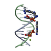 4i9vC  4pwmC  436dS S: Starting model for refinement C: citing same article ( |
|---|---|
| Similar structure data |


-Links
 PDBj
PDBj


- Assembly
| Deposited unit | 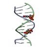
| ||||||||
|---|---|---|---|---|---|---|---|---|---|
| 1 |
| ||||||||
| Unit cell |
|
-Components
| #1: DNA chain | Mass: 3691.402 Da / Num. of mol.: 2 / Source method: obtained synthetically / Details: Chemically synthesized nucleic acid. #2: Water | ChemComp-HOH / |  Mass: 18.015 Da / Num. of mol.: 17 / Source method: isolated from a natural source / Formula: H2O Mass: 18.015 Da / Num. of mol.: 17 / Source method: isolated from a natural source / Formula: H2O |
|---|
-Experimental details
-Experiment
| Experiment | Method: X-RAY DIFFRACTION / Number of used crystals: 1 |
|---|
- Sample preparation
| Crystal | Density Matthews: 2.32 Å3/Da / Density % sol: 46.88 % |
|---|---|
| Crystal grow | Temperature: 291 K / Method: vapor diffusion, hanging drop / pH: 6 Details: 5% MPD, 20 mM Na Cacodylate, 6 mM Spermine tetra-HCl, 6 mM NaCl, 40 mM KCl , pH 6.0, VAPOR DIFFUSION, HANGING DROP, temperature 291K |
-Data collection
| Diffraction | Mean temperature: 100 K |
|---|---|
| Diffraction source | Source: SYNCHROTRON / Site: APS  / Beamline: 21-ID-D / Wavelength: 1 Å / Beamline: 21-ID-D / Wavelength: 1 Å |
| Detector | Type: MAR scanner 300 mm plate / Detector: IMAGE PLATE / Date: Apr 10, 2014 |
| Radiation | Monochromator: Si(111) / Protocol: SINGLE WAVELENGTH / Monochromatic (M) / Laue (L): M / Scattering type: x-ray |
| Radiation wavelength | Wavelength: 1 Å / Relative weight: 1 |
| Reflection | Resolution: 1.9→50 Å / Num. all: 5842 / Num. obs: 5801 / % possible obs: 99.3 % / Redundancy: 10.2 % / Rmerge(I) obs: 0.064 / Net I/σ(I): 60 |
| Reflection shell | Resolution: 1.9→1.93 Å / Redundancy: 6.7 % / Rmerge(I) obs: 0.738 / Mean I/σ(I) obs: 3.25 / % possible all: 100 |
- Processing
| Software |
| |||||||||||||||||||||||||||||||||||||||||||||||||||||||||||||||||
|---|---|---|---|---|---|---|---|---|---|---|---|---|---|---|---|---|---|---|---|---|---|---|---|---|---|---|---|---|---|---|---|---|---|---|---|---|---|---|---|---|---|---|---|---|---|---|---|---|---|---|---|---|---|---|---|---|---|---|---|---|---|---|---|---|---|---|
| Refinement | Method to determine structure: MOLECULAR REPLACEMENT Starting model: PDB ID 436D Resolution: 1.9→35.07 Å / Cor.coef. Fo:Fc: 0.97 / Cor.coef. Fo:Fc free: 0.967 / SU B: 4.094 / SU ML: 0.117 / Cross valid method: THROUGHOUT / σ(I): 5 / ESU R: 0.172 / ESU R Free: 0.147 / Stereochemistry target values: MAXIMUM LIKELIHOOD
| |||||||||||||||||||||||||||||||||||||||||||||||||||||||||||||||||
| Solvent computation | Ion probe radii: 0.8 Å / Shrinkage radii: 0.8 Å / VDW probe radii: 1.2 Å / Solvent model: MASK | |||||||||||||||||||||||||||||||||||||||||||||||||||||||||||||||||
| Refinement step | Cycle: LAST / Resolution: 1.9→35.07 Å
| |||||||||||||||||||||||||||||||||||||||||||||||||||||||||||||||||
| Refine LS restraints |
| |||||||||||||||||||||||||||||||||||||||||||||||||||||||||||||||||
| LS refinement shell | Resolution: 1.897→1.947 Å / Total num. of bins used: 20
|
