 Movie
Movie Controller

[English] 日本語
 Yorodumi
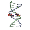
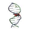
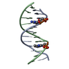
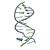
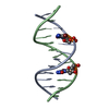
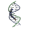
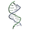
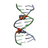
- PDB-4f3u: Crystal structure of 5-hydroxy-2'-deoxycytidine base paired with ... -
+ Open data

- Basic information
| Entry | Database: PDB / ID: 4f3u | ||||||||||||||||||||
|---|---|---|---|---|---|---|---|---|---|---|---|---|---|---|---|---|---|---|---|---|---|
| Title | Crystal structure of 5-hydroxy-2'-deoxycytidine base paired with 2'-deoxyguanosine in Dickerson Drew Dodecamer | ||||||||||||||||||||
 Components Components | DNA (5'-D(* Keywords KeywordsDNA / 5-hydroxy-dC / modified Dickerson / 5-hydroxy-2'-deoxycytidine | Function / homology | DNA / DNA (> 10) |  Function and homology information Function and homology informationBiological species | synthetic (others) | Method |  X-RAY DIFFRACTION / SYNCHROTRON / MOLECULAR REPLACEMENT / Resolution: 1.401 Å X-RAY DIFFRACTION / SYNCHROTRON / MOLECULAR REPLACEMENT / Resolution: 1.401 Å  Authors AuthorsSzulik, M.W. / Nocek, B. / Joachimiak, A. / Stone, M.P. |  CitationJournal: TO BE PUBLISHED CitationJournal: TO BE PUBLISHEDTitle: Crystal structure of 5-hydroxy-2'-deoxycytidine base paired with 2'-deoxyguanosine in Dickerson Drew Dodecamer Authors: Szulik, M.W. / Nocek, B. / Joachimiak, A. / Stone, M.P. History |
|
- Structure visualization
| Structure viewer | Molecule: MolmilJmol/JSmol |
|---|
- Downloads & links
-Download
| PDBx/mmCIF format | 4f3u.cif.gz | 40.8 KB | Display | PDBx/mmCIF format |
|---|---|---|---|---|
| PDB format | pdb4f3u.ent.gz | 28.9 KB | Display | PDB format |
| PDBx/mmJSON format | 4f3u.json.gz | Tree view | PDBx/mmJSON format | |
| Others |  Other downloads Other downloads |
-Validation report
| Arichive directory | https://data.pdbj.org/pub/pdb/validation_reports/f3/4f3uftp://data.pdbj.org/pub/pdb/validation_reports/f3/4f3u | HTTPS FTP |
|---|
-Related structure data
| Related structure data |  436dS S: Starting model for refinement |
|---|---|
| Similar structure data |


-Links
 PDBj
PDBj


- Assembly
| Deposited unit | 
| ||||||||
|---|---|---|---|---|---|---|---|---|---|
| 1 |
| ||||||||
| Unit cell |
|
-Components
| #1: DNA chain | Mass: 3679.392 Da / Num. of mol.: 2 / Source method: obtained synthetically / Source: (synth.) synthetic (others) #2: Chemical | ChemComp-MG / |   Mass: 24.305 Da / Num. of mol.: 1 / Source method: obtained synthetically / Formula: Mg Mass: 24.305 Da / Num. of mol.: 1 / Source method: obtained synthetically / Formula: Mg#3: Water | ChemComp-HOH / |  Mass: 18.015 Da / Num. of mol.: 94 / Source method: isolated from a natural source / Formula: H2O Mass: 18.015 Da / Num. of mol.: 94 / Source method: isolated from a natural source / Formula: H2O |
|---|
-Experimental details
-Experiment
| Experiment | Method: X-RAY DIFFRACTION / Number of used crystals: 1 |
|---|
- Sample preparation
| Crystal | Density Matthews: 2.26 Å3/Da / Density % sol: 45.62 % |
|---|---|
| Crystal grow | Temperature: 291 K / Method: vapor diffusion, hanging drop / pH: 7 Details: 10% MPD, 40mM sodium cacodylate, 12 mM spermine-tetra-HCl, 80mM sodium chloride, 20mM magnesium chloride., pH 7.0, VAPOR DIFFUSION, HANGING DROP, temperature 291K |
-Data collection
| Diffraction | Mean temperature: 100 K |
|---|---|
| Diffraction source | Source: SYNCHROTRON / Site: APS  / Beamline: 19-ID / Wavelength: 0.9794 Å / Beamline: 19-ID / Wavelength: 0.9794 Å |
| Detector | Type: ADSC QUANTUM 315r / Detector: CCD / Date: Feb 5, 2012 / Details: mirrors |
| Radiation | Monochromator: double crystal / Protocol: SINGLE WAVELENGTH / Monochromatic (M) / Laue (L): M / Scattering type: x-ray |
| Radiation wavelength | Wavelength: 0.9794 Å / Relative weight: 1 |
| Reflection | Resolution: 1.4→25 Å / Num. all: 13718 / Num. obs: 13485 / % possible obs: 98.3 % / Observed criterion σ(F): 2 / Redundancy: 6.7 % / Rmerge(I) obs: 0.051 / Net I/σ(I): 35.1 |
| Reflection shell | Resolution: 1.4→1.42 Å / % possible all: 95.4 |
- Processing
| Software |
| |||||||||||||||||||||||||||||||||||||||||||||||||||||||
|---|---|---|---|---|---|---|---|---|---|---|---|---|---|---|---|---|---|---|---|---|---|---|---|---|---|---|---|---|---|---|---|---|---|---|---|---|---|---|---|---|---|---|---|---|---|---|---|---|---|---|---|---|---|---|---|---|
| Refinement | Method to determine structure: MOLECULAR REPLACEMENT Starting model: PDB ENTRY 436D Resolution: 1.401→25 Å / Cor.coef. Fo:Fc: 0.97 / Cor.coef. Fo:Fc free: 0.956 / SU B: 3.612 / SU ML: 0.06 / Cross valid method: THROUGHOUT / ESU R: 0.069 / ESU R Free: 0.071 / Stereochemistry target values: MAXIMUM LIKELIHOOD / Details: HYDROGENS HAVE BEEN ADDED IN THE RIDING POSITIONS
| |||||||||||||||||||||||||||||||||||||||||||||||||||||||
| Solvent computation | Ion probe radii: 0.8 Å / Shrinkage radii: 0.8 Å / VDW probe radii: 1.2 Å / Solvent model: MASK | |||||||||||||||||||||||||||||||||||||||||||||||||||||||
| Displacement parameters | Biso mean: 27 Å2
| |||||||||||||||||||||||||||||||||||||||||||||||||||||||
| Refinement step | Cycle: LAST / Resolution: 1.401→25 Å
| |||||||||||||||||||||||||||||||||||||||||||||||||||||||
| Refine LS restraints |
| |||||||||||||||||||||||||||||||||||||||||||||||||||||||
| LS refinement shell | Resolution: 1.401→1.438 Å / Total num. of bins used: 20
|
