 Movie
Movie Controller

[English] 日本語
 Yorodumi
- PDB-263d: ISOHELICITY AND PHASING IN DRUG-DNA SEQUENCE RECOGNITION: CRYSTAL... -
+ Open data

- Basic information
| Entry | Database: PDB / ID: 263d | ||||||||||||||||||
|---|---|---|---|---|---|---|---|---|---|---|---|---|---|---|---|---|---|---|---|
| Title | ISOHELICITY AND PHASING IN DRUG-DNA SEQUENCE RECOGNITION: CRYSTAL STRUCTURE OF A TRIS(BENZIMIDAZOLE)-OLIGONUCLEOTIDE COMPLEX | ||||||||||||||||||
 Components Components | DNA (5'-D(* Keywords KeywordsDNA / B-DNA / DOUBLE HELIX / COMPLEXED WITH DRUG | Function / homology | Chem-TBZ / DNA / DNA (> 10) |  Function and homology information Function and homology informationMethod |  X-RAY DIFFRACTION / MOLECULAR REPLACEMENT / Resolution: 2.2 Å X-RAY DIFFRACTION / MOLECULAR REPLACEMENT / Resolution: 2.2 Å  Authors AuthorsClark, G.R. / Gray, E.J. / Neidle, S. / Li, Y.-H. / Leupin, W. |  CitationJournal: Biochemistry / Year: 1996 CitationJournal: Biochemistry / Year: 1996Title: Isohelicity and phasing in drug--DNA sequence recognition: crystal structure of a tris(benzimidazole)--oligonucleotide complex. Authors: Clark, G.R. / Gray, E.J. / Neidle, S. / Li, Y.H. / Leupin, W. #1: Journal: Nucleic Acids Res. / Year: 1994Title: Sequence-Dependent Effects in Drug-DNA Interaction: The Crystal Structure of Hoechst 33258 Bound to the d(CGCAAATTTGCG)2 Duplex Authors: Spink, N. / Skelly, J.V. / Neidle, S. #2: Journal: Eur.J.Biochem. / Year: 1994Title: Three-Dimensional Crystal Structure of the A-Tract DNA Dodecamer d(CGCAAATTTGCG) Complexed with the Minor-Groove-Binding Drug Hoechst 33258 Authors: Vega, M.C. / Saez, I.G. / Aymami, J. / Eritja, R. / van der Marel, G.A. / van Boom, J.H. / Rich, A. / Coll, M. History |
|
- Structure visualization
| Structure viewer | Molecule: MolmilJmol/JSmol |
|---|
- Downloads & links
-Download
| PDBx/mmCIF format | 263d.cif.gz | 25 KB | Display | PDBx/mmCIF format |
|---|---|---|---|---|
| PDB format | pdb263d.ent.gz | 16.2 KB | Display | PDB format |
| PDBx/mmJSON format | 263d.json.gz | Tree view | PDBx/mmJSON format | |
| Others |  Other downloads Other downloads |
-Validation report
| Arichive directory | https://data.pdbj.org/pub/pdb/validation_reports/63/263dftp://data.pdbj.org/pub/pdb/validation_reports/63/263d | HTTPS FTP |
|---|
-Related structure data
| Similar structure data |
|---|


-Links
 PDBj
PDBj


- Assembly
| Deposited unit | 
| ||||||||
|---|---|---|---|---|---|---|---|---|---|
| 1 |
| ||||||||
| Unit cell |
|
-Components
| #1: DNA chain | Mass: 3662.404 Da / Num. of mol.: 2 / Source method: obtained synthetically #2: Chemical | ChemComp-TBZ / | 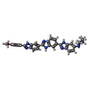  Mass: 541.626 Da / Num. of mol.: 1 / Source method: obtained synthetically / Formula: C32H29N8O Mass: 541.626 Da / Num. of mol.: 1 / Source method: obtained synthetically / Formula: C32H29N8O#3: Water | ChemComp-HOH / |  Mass: 18.015 Da / Num. of mol.: 61 / Source method: isolated from a natural source / Formula: H2O Mass: 18.015 Da / Num. of mol.: 61 / Source method: isolated from a natural source / Formula: H2O |
|---|
-Experimental details
-Experiment
| Experiment | Method: X-RAY DIFFRACTION / Number of used crystals: 1 |
|---|
- Sample preparation
| Crystal | Density Matthews: 2.23 Å3/Da / Density % sol: 44.93 % | ||||||||||||||||||||||||||||||||||||||||||||||||
|---|---|---|---|---|---|---|---|---|---|---|---|---|---|---|---|---|---|---|---|---|---|---|---|---|---|---|---|---|---|---|---|---|---|---|---|---|---|---|---|---|---|---|---|---|---|---|---|---|---|
| Crystal grow | Temperature: 286 K / Method: vapor diffusion, hanging drop / pH: 7 Details: pH 7.00, VAPOR DIFFUSION, HANGING DROP, temperature 286.00K | ||||||||||||||||||||||||||||||||||||||||||||||||
| Components of the solutions |
| ||||||||||||||||||||||||||||||||||||||||||||||||
| Crystal grow | *PLUS pH: 7 | ||||||||||||||||||||||||||||||||||||||||||||||||
| Components of the solutions | *PLUS
|
-Data collection
| Diffraction | Mean temperature: 287 K |
|---|---|
| Diffraction source | Source: ROTATING ANODE / Wavelength: 1.5418 |
| Detector | Type: XENTRONICS / Detector: AREA DETECTOR / Date: May 15, 1996 |
| Radiation | Monochromator: GRAPHITE / Monochromatic (M) / Laue (L): M / Scattering type: x-ray |
| Radiation wavelength | Wavelength: 1.5418 Å / Relative weight: 1 |
| Reflection | Resolution: 2.2→8 Å / Num. obs: 3594 / % possible obs: 92 % / Observed criterion σ(I): 2 / Rmerge(I) obs: 0.083 |
| Reflection shell | Resolution: 2.2→2.35 Å / % possible all: 57 |
| Reflection | *PLUS Highest resolution: 2.2 Å / Lowest resolution: 8 Å / % possible obs: 92 % / Observed criterion σ(I): 2 |
| Reflection shell | *PLUS Highest resolution: 2.2 Å / Lowest resolution: 2.35 Å / % possible obs: 57 % |
- Processing
| Software |
| ||||||||||||||||||||||||||||||||||||||||||||||||||||||||||||
|---|---|---|---|---|---|---|---|---|---|---|---|---|---|---|---|---|---|---|---|---|---|---|---|---|---|---|---|---|---|---|---|---|---|---|---|---|---|---|---|---|---|---|---|---|---|---|---|---|---|---|---|---|---|---|---|---|---|---|---|---|---|
| Refinement | Method to determine structure: MOLECULAR REPLACEMENT Starting model: GDL023 Resolution: 2.2→8 Å / σ(F): 2
| ||||||||||||||||||||||||||||||||||||||||||||||||||||||||||||
| Displacement parameters | Biso mean: 24 Å2 | ||||||||||||||||||||||||||||||||||||||||||||||||||||||||||||
| Refine Biso | Class: ALL ATOMS / Details: TR / Treatment: isotropic | ||||||||||||||||||||||||||||||||||||||||||||||||||||||||||||
| Refinement step | Cycle: LAST / Resolution: 2.2→8 Å
| ||||||||||||||||||||||||||||||||||||||||||||||||||||||||||||
| Refine LS restraints |
| ||||||||||||||||||||||||||||||||||||||||||||||||||||||||||||
| Software | *PLUS Name: X-PLOR / Classification: refinement | ||||||||||||||||||||||||||||||||||||||||||||||||||||||||||||
| Refinement | *PLUS Highest resolution: 2.2 Å / Lowest resolution: 8 Å / σ(F): 2 | ||||||||||||||||||||||||||||||||||||||||||||||||||||||||||||
| Solvent computation | *PLUS | ||||||||||||||||||||||||||||||||||||||||||||||||||||||||||||
| Displacement parameters | *PLUS |
