 Movie
Movie Controller

[English] 日本語
 Yorodumi
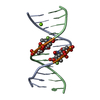
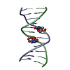
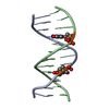
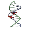
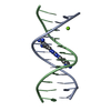
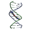
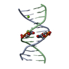
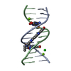
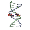
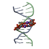
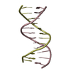
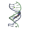
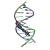
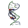
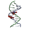
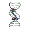
- PDB-1g75: MOLECULAR AND CRYSTAL STRUCTURE OF D(CGCGAATF5UCGCG): 5-FORMYLURI... -
+ Open data

- Basic information
| Entry | Database: PDB / ID: 1g75 | ||||||||||||||||||
|---|---|---|---|---|---|---|---|---|---|---|---|---|---|---|---|---|---|---|---|
| Title | MOLECULAR AND CRYSTAL STRUCTURE OF D(CGCGAATF5UCGCG): 5-FORMYLURIDINE/ ADENOSINE BASE-PAIRS IN B-DNA | ||||||||||||||||||
 Components Components | 5'-D(* Keywords KeywordsDNA / MODIFIED NUCLEOTIDE / FORMYLURIDINE / DNA DAMAGE / B-DNA / DOUBLE HELIX / DEOXYRIBONUCLEIC ACID | Function / homology | : / DNA / DNA (> 10) |  Function and homology information Function and homology informationMethod |  X-RAY DIFFRACTION / SYNCHROTRON / MOLECULAR REPLACEMENT / Resolution: 1.57 Å X-RAY DIFFRACTION / SYNCHROTRON / MOLECULAR REPLACEMENT / Resolution: 1.57 Å  Authors AuthorsTsunoda, M. / Karino, N. / Ueno, Y. / Matsuda, A. / Takenaka, A. |  CitationJournal: Acta Crystallogr.,Sect.D / Year: 2001 CitationJournal: Acta Crystallogr.,Sect.D / Year: 2001Title: Crystallization and preliminary X-ray analysis of a DNA dodecamer containing 2'-deoxy-5-formyluridine; what is the role of magnesium cation in crystallization of Dickerson-type DNA dodecamers? Authors: Tsunoda, M. / Karino, N. / Ueno, Y. / Matsuda, A. / Takenaka, A. History |
|
- Structure visualization
| Structure viewer | Molecule: MolmilJmol/JSmol |
|---|
- Downloads & links
-Download
| PDBx/mmCIF format | 1g75.cif.gz | 27.8 KB | Display | PDBx/mmCIF format |
|---|---|---|---|---|
| PDB format | pdb1g75.ent.gz | 17.9 KB | Display | PDB format |
| PDBx/mmJSON format | 1g75.json.gz | Tree view | PDBx/mmJSON format | |
| Others |  Other downloads Other downloads |
-Validation report
| Arichive directory | https://data.pdbj.org/pub/pdb/validation_reports/g7/1g75ftp://data.pdbj.org/pub/pdb/validation_reports/g7/1g75 | HTTPS FTP |
|---|
-Related structure data
| Related structure data | 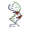 1g8nC  1g8uC  1g8vC 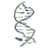 355dS S: Starting model for refinement C: citing same article ( |
|---|---|
| Similar structure data | |
| Other databases |
|


-Links
 PDBj
PDBj


- Assembly
| Deposited unit | 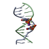
| ||||||||
|---|---|---|---|---|---|---|---|---|---|
| 1 |
| ||||||||
| Unit cell |
|
-Components
| #1: DNA chain | Mass: 3677.376 Da / Num. of mol.: 2 / Source method: obtained synthetically #2: Chemical | ChemComp-K / |   Mass: 39.098 Da / Num. of mol.: 1 / Source method: obtained synthetically / Formula: K Mass: 39.098 Da / Num. of mol.: 1 / Source method: obtained synthetically / Formula: K#3: Chemical | ChemComp-MG / |   Mass: 24.305 Da / Num. of mol.: 1 / Source method: obtained synthetically / Formula: Mg Mass: 24.305 Da / Num. of mol.: 1 / Source method: obtained synthetically / Formula: Mg#4: Water | ChemComp-HOH / |  Mass: 18.015 Da / Num. of mol.: 150 / Source method: isolated from a natural source / Formula: H2O Mass: 18.015 Da / Num. of mol.: 150 / Source method: isolated from a natural source / Formula: H2O |
|---|
-Experimental details
-Experiment
| Experiment | Method: X-RAY DIFFRACTION / Number of used crystals: 1 |
|---|
- Sample preparation
| Crystal | Density Matthews: 2.15 Å3/Da / Density % sol: 42.89 % | ||||||||||||||||||||||||||||||||||||||||||||||||||||||
|---|---|---|---|---|---|---|---|---|---|---|---|---|---|---|---|---|---|---|---|---|---|---|---|---|---|---|---|---|---|---|---|---|---|---|---|---|---|---|---|---|---|---|---|---|---|---|---|---|---|---|---|---|---|---|---|
| Crystal grow | Temperature: 277 K / Method: vapor diffusion, sitting drop / pH: 6.5 Details: MPD, spermine, magnesium chloride, potassium chloride, sodium chloride, cacodylate, pH 6.5, VAPOR DIFFUSION, SITTING DROP, temperature 277K | ||||||||||||||||||||||||||||||||||||||||||||||||||||||
| Components of the solutions |
| ||||||||||||||||||||||||||||||||||||||||||||||||||||||
| Crystal grow | *PLUS Method: vapor diffusion, hanging drop | ||||||||||||||||||||||||||||||||||||||||||||||||||||||
| Components of the solutions | *PLUS
|
-Data collection
| Diffraction | Mean temperature: 100 K |
|---|---|
| Diffraction source | Source: SYNCHROTRON / Site: Photon Factory  / Beamline: BL-18B / Wavelength: 1 Å / Beamline: BL-18B / Wavelength: 1 Å |
| Detector | Type: WEISSENBERG / Detector: DIFFRACTOMETER / Date: Jun 3, 2000 |
| Radiation | Protocol: SINGLE WAVELENGTH / Monochromatic (M) / Laue (L): M / Scattering type: x-ray |
| Radiation wavelength | Wavelength: 1 Å / Relative weight: 1 |
| Reflection | Resolution: 1.57→50 Å / Num. all: 90663 / Num. obs: 9502 / % possible obs: 97.1 % / Observed criterion σ(I): -3 / Redundancy: 9.54 % / Rmerge(I) obs: 0.031 |
| Reflection shell | Resolution: 1.57→1.6 Å / Rmerge(I) obs: 0.266 / % possible all: 89.9 |
| Reflection | *PLUS Num. measured all: 90663 |
- Processing
| Software |
| ||||||||||||||||||
|---|---|---|---|---|---|---|---|---|---|---|---|---|---|---|---|---|---|---|---|
| Refinement | Method to determine structure: MOLECULAR REPLACEMENT Starting model: PDB ENTRY 355D Resolution: 1.57→50 Å / Cross valid method: THROUGHOUT / σ(F): 0
| ||||||||||||||||||
| Refine analyze |
| ||||||||||||||||||
| Refinement step | Cycle: LAST / Resolution: 1.57→50 Å
| ||||||||||||||||||
| Refine LS restraints |
| ||||||||||||||||||
| LS refinement shell | Resolution: 1.57→1.67 Å / Rfactor Rfree error: 0.021 /
| ||||||||||||||||||
| Software | *PLUS Name: CNS / Version: 1 / Classification: refinement | ||||||||||||||||||
| Refinement | *PLUS Lowest resolution: 50 Å / σ(F): 0 / Rfactor obs: 0.21 / Rfactor Rwork: 0.21 / % reflection Rfree: 10 % | ||||||||||||||||||
| Solvent computation | *PLUS | ||||||||||||||||||
| Displacement parameters | *PLUS | ||||||||||||||||||
| Refine LS restraints | *PLUS
| ||||||||||||||||||
| LS refinement shell | *PLUS Rfactor Rfree: 0.263 / Rfactor Rwork: 0.235 |
