 Movie
Movie Controller

[English] 日本語
 Yorodumi
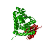
- PDB-4q3n: Crystal structure of MGS-M5, a lactate dehydrogenase enzyme from ... -
+ Open data

- Basic information
| Entry | Database: PDB / ID: 4q3n | ||||||
|---|---|---|---|---|---|---|---|
| Title | Crystal structure of MGS-M5, a lactate dehydrogenase enzyme from a Medee basin deep-sea metagenome library | ||||||
 Components Components | MGS-M5 | ||||||
 Keywords Keywords | HYDROLASE / metagenome / metagenomic library / ROSSMANN FOLD / DEHYDROGENASE / OXIDOREDUCTASE / L-LACTATE DEHYDROGENASE | ||||||
| Function / homology |  Function and homology information Function and homology informationL-lactate dehydrogenase / L-lactate dehydrogenase (NAD+) activity / lactate metabolic process / glycolytic process / cytoplasm Similarity search - Function | ||||||
| Biological species | unidentified (others) | ||||||
| Method |  X-RAY DIFFRACTION / MOLECULAR REPLACEMENT / Resolution: 1.97 Å X-RAY DIFFRACTION / MOLECULAR REPLACEMENT / Resolution: 1.97 Å | ||||||
 Authors Authors | Stogios, P.J. / Xu, X. / Cui, H. / Alcaide, M. / Ferrer, M. / Savchenko, A. | ||||||
 Citation Citation | Journal: Environ Microbiol / Year: 2015 Title: Pressure adaptation is linked to thermal adaptation in salt-saturated marine habitats. Authors: Alcaide, M. / Stogios, P.J. / Lafraya, A. / Tchigvintsev, A. / Flick, R. / Bargiela, R. / Chernikova, T.N. / Reva, O.N. / Hai, T. / Leggewie, C.C. / Katzke, N. / La Cono, V. / Matesanz, R. / ...Authors: Alcaide, M. / Stogios, P.J. / Lafraya, A. / Tchigvintsev, A. / Flick, R. / Bargiela, R. / Chernikova, T.N. / Reva, O.N. / Hai, T. / Leggewie, C.C. / Katzke, N. / La Cono, V. / Matesanz, R. / Jebbar, M. / Jaeger, K.E. / Yakimov, M.M. / Yakunin, A.F. / Golyshin, P.N. / Golyshina, O.V. / Savchenko, A. / Ferrer, M. | ||||||
| History |
|
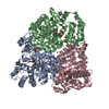
- Structure visualization
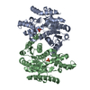
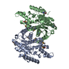
| Structure viewer | Molecule: MolmilJmol/JSmol |
|---|
- Downloads & links
-Download
| PDBx/mmCIF format | 4q3n.cif.gz | 150.5 KB | Display | PDBx/mmCIF format |
|---|---|---|---|---|
| PDB format | pdb4q3n.ent.gz | 118.1 KB | Display | PDB format |
| PDBx/mmJSON format | 4q3n.json.gz | Tree view | PDBx/mmJSON format | |
| Others |  Other downloads Other downloads |
-Validation report
| Arichive directory | https://data.pdbj.org/pub/pdb/validation_reports/q3/4q3nftp://data.pdbj.org/pub/pdb/validation_reports/q3/4q3n | HTTPS FTP |
|---|
-Related structure data
| Related structure data |  4q3kC  4q3lC  4q3mC  4q3oC  1ldnS C: citing same article ( S: Starting model for refinement |
|---|---|
| Similar structure data |

-Links
 PDBj
PDBj


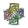
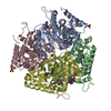
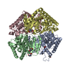
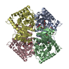
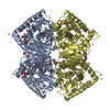
- Assembly
| Deposited unit | 
| ||||||||
|---|---|---|---|---|---|---|---|---|---|
| 1 | 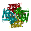
| ||||||||
| Unit cell |
| ||||||||
| Components on special symmetry positions |
|
-Components
-Protein , 1 types, 1 molecules A
| #1: Protein | Mass: 34817.762 Da / Num. of mol.: 1 Source method: isolated from a genetically manipulated source Source: (gene. exp.) unidentified (others) / Gene: MGS-M5 / Plasmid: p15-TV LIC / Production host:  |
|---|
-Non-polymers , 7 types, 370 molecules 


| #2: Chemical | ChemComp-NA /  Mass: 22.990 Da / Num. of mol.: 1 / Source method: obtained synthetically / Formula: Na Mass: 22.990 Da / Num. of mol.: 1 / Source method: obtained synthetically / Formula: Na | ||||||||||
|---|---|---|---|---|---|---|---|---|---|---|---|
| #3: Chemical |  Mass: 35.453 Da / Num. of mol.: 2 / Source method: obtained synthetically / Formula: Cl Mass: 35.453 Da / Num. of mol.: 2 / Source method: obtained synthetically / Formula: Cl#4: Chemical | ChemComp-ACT /  Mass: 59.044 Da / Num. of mol.: 6 / Source method: obtained synthetically / Formula: C2H3O2 Mass: 59.044 Da / Num. of mol.: 6 / Source method: obtained synthetically / Formula: C2H3O2#5: Chemical |  Mass: 150.173 Da / Num. of mol.: 2 / Source method: obtained synthetically / Formula: C6H14O4 Mass: 150.173 Da / Num. of mol.: 2 / Source method: obtained synthetically / Formula: C6H14O4#6: Chemical | ChemComp-GOL /  Mass: 92.094 Da / Num. of mol.: 5 / Source method: obtained synthetically / Formula: C3H8O3 Mass: 92.094 Da / Num. of mol.: 5 / Source method: obtained synthetically / Formula: C3H8O3#7: Chemical | ChemComp-TRS / |  Mass: 122.143 Da / Num. of mol.: 1 / Source method: obtained synthetically / Formula: C4H12NO3 / Comment: pH buffer*YM Mass: 122.143 Da / Num. of mol.: 1 / Source method: obtained synthetically / Formula: C4H12NO3 / Comment: pH buffer*YM#8: Water | ChemComp-HOH / | Mass: 18.015 Da / Num. of mol.: 353 / Source method: isolated from a natural source / Formula: H2O |
-Details
| Source details | THE SAMPLE HAS BEEN OBTAINED FROM LAKE MATAPAN DEEP-SEA AND IDENTIFIED |
|---|
-Experimental details
-Experiment
| Experiment | Method: X-RAY DIFFRACTION / Number of used crystals: 1 |
|---|
- Sample preparation
| Crystal | Density Matthews: 3.15 Å3/Da / Density % sol: 60.97 % |
|---|---|
| Crystal grow | Temperature: 287 K / Method: vapor diffusion, hanging drop / pH: 6.5 Details: 0.1 M sodium cacodylate, 0.2 M calcium acetate, 9% PEG 8K, trypsin in 1/70 molar ratio protein:trypsin cryoprotectant: 12% glycerol then paratone, pH 6.5, VAPOR DIFFUSION, HANGING DROP, temperature 287K |
-Data collection
| Diffraction | Mean temperature: 100 K |
|---|---|
| Diffraction source | Source: ROTATING ANODE / Type: RIGAKU MICROMAX-007 HF / Wavelength: 1.5418 Å |
| Detector | Type: RIGAKU RAXIS IV++ / Detector: IMAGE PLATE / Date: Nov 6, 2012 |
| Radiation | Monochromator: GRAPHITE / Protocol: SINGLE WAVELENGTH / Monochromatic (M) / Laue (L): M / Scattering type: x-ray |
| Radiation wavelength | Wavelength: 1.5418 Å / Relative weight: 1 |
| Reflection | Resolution: 1.97→35 Å / Num. obs: 31973 / % possible obs: 99.9 % / Observed criterion σ(F): 0 / Observed criterion σ(I): -2 / Redundancy: 6.1 % / Rmerge(I) obs: 0.048 / Net I/σ(I): 45.3 |
| Reflection shell | Resolution: 1.97→2 Å / Redundancy: 6 % / Rmerge(I) obs: 0.51 / Mean I/σ(I) obs: 5.31 / % possible all: 99.9 |
- Processing
| Software |
| |||||||||||||||||||||||||||||||||||||||||||||||||||||||||||||||||||||||||||||||||||||||||||||||||||||||||
|---|---|---|---|---|---|---|---|---|---|---|---|---|---|---|---|---|---|---|---|---|---|---|---|---|---|---|---|---|---|---|---|---|---|---|---|---|---|---|---|---|---|---|---|---|---|---|---|---|---|---|---|---|---|---|---|---|---|---|---|---|---|---|---|---|---|---|---|---|---|---|---|---|---|---|---|---|---|---|---|---|---|---|---|---|---|---|---|---|---|---|---|---|---|---|---|---|---|---|---|---|---|---|---|---|---|---|
| Refinement | Method to determine structure: MOLECULAR REPLACEMENT Starting model: PDB entry 1LDN Resolution: 1.97→34.31 Å / SU ML: 0.19 / σ(F): 1.34 / Phase error: 18.77 / Stereochemistry target values: ML
| |||||||||||||||||||||||||||||||||||||||||||||||||||||||||||||||||||||||||||||||||||||||||||||||||||||||||
| Solvent computation | Shrinkage radii: 0.9 Å / VDW probe radii: 1.11 Å / Solvent model: FLAT BULK SOLVENT MODEL | |||||||||||||||||||||||||||||||||||||||||||||||||||||||||||||||||||||||||||||||||||||||||||||||||||||||||
| Refinement step | Cycle: LAST / Resolution: 1.97→34.31 Å
| |||||||||||||||||||||||||||||||||||||||||||||||||||||||||||||||||||||||||||||||||||||||||||||||||||||||||
| Refine LS restraints |
| |||||||||||||||||||||||||||||||||||||||||||||||||||||||||||||||||||||||||||||||||||||||||||||||||||||||||
| LS refinement shell |
| |||||||||||||||||||||||||||||||||||||||||||||||||||||||||||||||||||||||||||||||||||||||||||||||||||||||||
| Refinement TLS params. | Method: refined / Refine-ID: X-RAY DIFFRACTION
| |||||||||||||||||||||||||||||||||||||||||||||||||||||||||||||||||||||||||||||||||||||||||||||||||||||||||
| Refinement TLS group |
|
