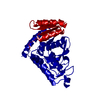
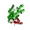
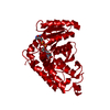
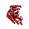
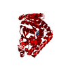
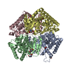
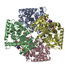
Entry Database : PDB / ID : 2x8lTitle Plasmodium falciparum lactate dehydrogenase apo structure L-LACTATE DEHYDROGENASE Keywords / / / Function / homology Function Domain/homology Component
/ / / / / / / / / / / / / / / / / / / / / / / / / / Biological species PLASMODIUM FALCIPARUM (malaria parasite P. falciparum)Method / / / Resolution : 1.6 Å Authors Birkinshaw, R.W. / Brady, R.L. Journal : To be Published Title : The Apo Crystal Structure of Plasmodium Falciparum Lactate DehydrogenaseAuthors : Birkinshaw, R.W. / Brady, R.L. History Deposition Mar 10, 2010 Deposition site / Processing site Revision 1.0 Mar 23, 2010 Provider / Type Revision 1.1 Jul 13, 2011 Group / Version format complianceRevision 1.2 Jan 30, 2019 Group Advisory / Data collection ... Advisory / Data collection / Derived calculations / Experimental preparation / Other Category exptl_crystal_grow / pdbx_database_proc ... exptl_crystal_grow / pdbx_database_proc / pdbx_database_status / pdbx_struct_special_symmetry / pdbx_unobs_or_zero_occ_atoms Item / _pdbx_database_status.recvd_author_approvalRevision 1.3 Feb 6, 2019 Group / Experimental preparation / Category / Item Revision 1.4 Dec 20, 2023 Group Advisory / Data collection ... Advisory / Data collection / Database references / Derived calculations / Other / Refinement description Category chem_comp_atom / chem_comp_bond ... chem_comp_atom / chem_comp_bond / database_2 / pdbx_database_status / pdbx_initial_refinement_model / pdbx_unobs_or_zero_occ_atoms / struct_site Item _database_2.pdbx_DOI / _database_2.pdbx_database_accession ... _database_2.pdbx_DOI / _database_2.pdbx_database_accession / _pdbx_database_status.status_code_sf / _struct_site.pdbx_auth_asym_id / _struct_site.pdbx_auth_comp_id / _struct_site.pdbx_auth_seq_id
Show all Show less
 Movie
Movie Controller


 Open data
 Basic information
 Components
 Keywords
 Function and homology information
 X-RAY DIFFRACTION /
  Authors
 Citation
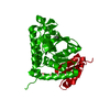
 Structure visualization
 Downloads & links
 Other downloads
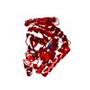


 PDBj
PDBj


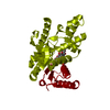
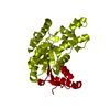
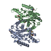
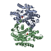
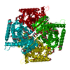
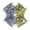
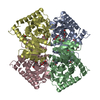
 Assembly


 Mass: 92.094 Da / Num. of mol.: 1 / Source method: obtained synthetically / Formula: C3H8O3
 Mass: 18.015 Da / Num. of mol.: 318 / Source method: isolated from a natural source / Formula: H2O
 Sample preparation
 / Beamline: I02 / Wavelength: 0.9795
  Processing