 Movie
Movie Controller

[English] 日本語
 Yorodumi
- PDB-2a94: Structure of Plasmodium falciparum lactate dehydrogenase complexe... -
+ Open data

- Basic information
| Entry | Database: PDB / ID: 2a94 | |||||||||
|---|---|---|---|---|---|---|---|---|---|---|
| Title | Structure of Plasmodium falciparum lactate dehydrogenase complexed to APADH. | |||||||||
 Components Components | L-lactate dehydrogenase | |||||||||
 Keywords Keywords | OXIDOREDUCTASE / Rossmann fold | |||||||||
| Function / homology |  Function and homology information Function and homology informationL-lactate dehydrogenase / L-lactate dehydrogenase (NAD+) activity / lactate metabolic process Similarity search - Function | |||||||||
| Biological species |  | |||||||||
| Method |  X-RAY DIFFRACTION / SYNCHROTRON / MOLECULAR REPLACEMENT / Resolution: 1.5 Å X-RAY DIFFRACTION / SYNCHROTRON / MOLECULAR REPLACEMENT / Resolution: 1.5 Å | |||||||||
 Authors Authors | Chaikuad, A. / Fairweather, V. / Conners, R. / Joseph-Horne, T. / Turgut-Balik, D. / Brady, R.L. | |||||||||
 Citation Citation | Journal: Biochemistry / Year: 2005 Title: Structure of Lactate Dehydrogenase from Plasmodium vivax: Complexes with NADH and APADH. Authors: Chaikuad, A. / Fairweather, V. / Conners, R. / Joseph-Horne, T. / Turgut-Balik, D. / Brady, R.L. | |||||||||
| History |
|
- Structure visualization
| Structure viewer | Molecule: MolmilJmol/JSmol |
|---|
- Downloads & links
-Download
| PDBx/mmCIF format | 2a94.cif.gz | 80.3 KB | Display | PDBx/mmCIF format |
|---|---|---|---|---|
| PDB format | pdb2a94.ent.gz | 58.2 KB | Display | PDB format |
| PDBx/mmJSON format | 2a94.json.gz | Tree view | PDBx/mmJSON format | |
| Others |  Other downloads Other downloads |
-Validation report
| Arichive directory | https://data.pdbj.org/pub/pdb/validation_reports/a9/2a94ftp://data.pdbj.org/pub/pdb/validation_reports/a9/2a94 | HTTPS FTP |
|---|
-Related structure data
| Related structure data |  2a92C  2aa3C  1t2cS S: Starting model for refinement C: citing same article ( |
|---|---|
| Similar structure data |


-Links
 PDBj
PDBj


- Assembly
| Deposited unit | 
| ||||||||
|---|---|---|---|---|---|---|---|---|---|
| 1 | 
| ||||||||
| Unit cell |
| ||||||||
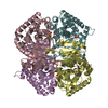
| Details | Biological unit is a tetramer |
-Components
| #1: Protein | Mass: 34846.387 Da / Num. of mol.: 1 Source method: isolated from a genetically manipulated source Source: (gene. exp.) Gene: LDH / Plasmid: pKK223-3 / Production host:  |
|---|---|
| #2: Chemical | ChemComp-AP0 /   Mass: 664.453 Da / Num. of mol.: 1 / Source method: obtained synthetically / Formula: C22H30N6O14P2 Mass: 664.453 Da / Num. of mol.: 1 / Source method: obtained synthetically / Formula: C22H30N6O14P2 |
| #3: Water | ChemComp-HOH /  Mass: 18.015 Da / Num. of mol.: 214 / Source method: isolated from a natural source / Formula: H2O Mass: 18.015 Da / Num. of mol.: 214 / Source method: isolated from a natural source / Formula: H2O |
-Experimental details
-Experiment
| Experiment | Method: X-RAY DIFFRACTION / Number of used crystals: 1 |
|---|
- Sample preparation
| Crystal | Density Matthews: 2.31 Å3/Da / Density % sol: 46.33 % |
|---|---|
| Crystal grow | Temperature: 291 K / Method: vapor diffusion, hanging drop / pH: 7.5 Details: MPD, pH 7.5, VAPOR DIFFUSION, HANGING DROP, temperature 291K |
-Data collection
| Diffraction | Mean temperature: 100 K |
|---|---|
| Diffraction source | Source: SYNCHROTRON / Site: SRS  / Beamline: PX14.2 / Wavelength: 0.978 / Wavelength: 0.978 Å / Beamline: PX14.2 / Wavelength: 0.978 / Wavelength: 0.978 Å |
| Detector | Type: ADSC QUANTUM 4 / Detector: CCD / Date: Jun 24, 2001 |
| Radiation | Monochromator: Si111 / Protocol: SINGLE WAVELENGTH / Monochromatic (M) / Laue (L): M / Scattering type: x-ray |
| Radiation wavelength | Wavelength: 0.978 Å / Relative weight: 1 |
| Reflection | Resolution: 1.5→30 Å / Num. all: 51103 / Num. obs: 50501 / % possible obs: 99.5 % / Observed criterion σ(F): 0 / Observed criterion σ(I): 2 / Redundancy: 4.5 % / Rmerge(I) obs: 0.031 / Χ2: 1.037 / Net I/σ(I): 37.4 |
| Reflection shell | Resolution: 1.5→1.62 Å / % possible obs: 99.5 % / Redundancy: 4.1 % / Rmerge(I) obs: 0.133 / Mean I/σ(I) obs: 7.3 / Num. measured obs: 10030 / Χ2: 0.778 / % possible all: 99.5 |
- Processing
| Software |
| |||||||||||||||||||||||||||||||||||||||||||||||||||||||||||||||||||||||||||||||||||||||||||||||||||||||||
|---|---|---|---|---|---|---|---|---|---|---|---|---|---|---|---|---|---|---|---|---|---|---|---|---|---|---|---|---|---|---|---|---|---|---|---|---|---|---|---|---|---|---|---|---|---|---|---|---|---|---|---|---|---|---|---|---|---|---|---|---|---|---|---|---|---|---|---|---|---|---|---|---|---|---|---|---|---|---|---|---|---|---|---|---|---|---|---|---|---|---|---|---|---|---|---|---|---|---|---|---|---|---|---|---|---|---|
| Refinement | Method to determine structure: MOLECULAR REPLACEMENT Starting model: PDB Entry 1T2C Resolution: 1.5→29.36 Å / Cor.coef. Fo:Fc: 0.963 / Cor.coef. Fo:Fc free: 0.957 / SU B: 1.473 / SU ML: 0.055 / Isotropic thermal model: Isotropic / Cross valid method: THROUGHOUT / σ(F): 0 / ESU R: 0.066 / ESU R Free: 0.064 / Stereochemistry target values: MAXIMUM LIKELIHOOD / Details: HYDROGENS HAVE BEEN ADDED IN THE RIDING POSITIONS
| |||||||||||||||||||||||||||||||||||||||||||||||||||||||||||||||||||||||||||||||||||||||||||||||||||||||||
| Solvent computation | Ion probe radii: 0.8 Å / Shrinkage radii: 0.8 Å / VDW probe radii: 1.4 Å / Solvent model: BABINET MODEL WITH MASK | |||||||||||||||||||||||||||||||||||||||||||||||||||||||||||||||||||||||||||||||||||||||||||||||||||||||||
| Displacement parameters | Biso mean: 10.543 Å2
| |||||||||||||||||||||||||||||||||||||||||||||||||||||||||||||||||||||||||||||||||||||||||||||||||||||||||
| Refinement step | Cycle: LAST / Resolution: 1.5→29.36 Å
| |||||||||||||||||||||||||||||||||||||||||||||||||||||||||||||||||||||||||||||||||||||||||||||||||||||||||
| Refine LS restraints |
| |||||||||||||||||||||||||||||||||||||||||||||||||||||||||||||||||||||||||||||||||||||||||||||||||||||||||
| LS refinement shell | Resolution: 1.5→1.539 Å / Total num. of bins used: 20
|
