 Movie
Movie Controller


+ Open data

- Basic information
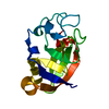
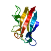
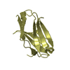
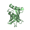
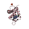
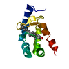
| Entry | Database: PDB / ID: 4pui | ||||||
|---|---|---|---|---|---|---|---|
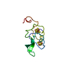
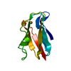
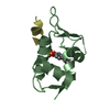
| Title | BolA domain of SufE1 from Arabidopsis thaliana | ||||||
 Components Components | SufE-like protein, chloroplastic | ||||||
 Keywords Keywords | PROTEIN BINDING / stress-responsive protein / METAL BINDING PROTEIN / PROTEIN BINDING PROTEIN | ||||||
| Function / homology |  Function and homology information Function and homology informationembryo development ending in seed dormancy / chloroplast stroma / chloroplast / enzyme activator activity / mitochondrion / cytosol Similarity search - Function | ||||||
| Biological species |  | ||||||
| Method |  X-RAY DIFFRACTION / SYNCHROTRON / MOLECULAR REPLACEMENT / Resolution: 1.698 Å X-RAY DIFFRACTION / SYNCHROTRON / MOLECULAR REPLACEMENT / Resolution: 1.698 Å | ||||||
 Authors Authors | Roret, T. / Didierjean, C. | ||||||
 Citation Citation | Journal: J.Biol.Chem. / Year: 2014 Title: Structural and Spectroscopic Insights into BolA-Glutaredoxin Complexes. Authors: Roret, T. / Tsan, P. / Couturier, J. / Zhang, B. / Johnson, M.K. / Rouhier, N. / Didierjean, C. | ||||||
| History |
|
- Structure visualization
| Structure viewer | Molecule: MolmilJmol/JSmol |
|---|
- Downloads & links
-Download
| PDBx/mmCIF format | 4pui.cif.gz | 83.7 KB | Display | PDBx/mmCIF format |
|---|---|---|---|---|
| PDB format | pdb4pui.ent.gz | 63.9 KB | Display | PDB format |
| PDBx/mmJSON format | 4pui.json.gz | Tree view | PDBx/mmJSON format | |
| Others |  Other downloads Other downloads |
-Validation report
| Arichive directory | https://data.pdbj.org/pub/pdb/validation_reports/pu/4puiftp://data.pdbj.org/pub/pdb/validation_reports/pu/4pui | HTTPS FTP |
|---|
-Related structure data
| Related structure data |  2mm9C  2mmaC 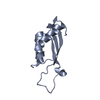 4pugC  4puhC  1v60S C: citing same article ( S: Starting model for refinement |
|---|---|
| Similar structure data |

-Links
 PDBj
PDBj- Assembly
| Deposited unit | 
| ||||||||
|---|---|---|---|---|---|---|---|---|---|
| 1 | 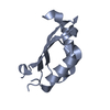
| ||||||||
| 2 | 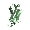
| ||||||||
| Unit cell |
|
-Components
| #1: Protein | Mass: 10639.063 Da / Num. of mol.: 2 / Fragment: UNP RESIDUES 277-371 Source method: isolated from a genetically manipulated source Source: (gene. exp.)  #2: Water | ChemComp-HOH / |  Mass: 18.015 Da / Num. of mol.: 165 / Source method: isolated from a natural source / Formula: H2O Mass: 18.015 Da / Num. of mol.: 165 / Source method: isolated from a natural source / Formula: H2O |
|---|
-Experimental details
-Experiment
| Experiment | Method: X-RAY DIFFRACTION / Number of used crystals: 1 |
|---|
- Sample preparation
| Crystal | Density Matthews: 2.18 Å3/Da / Density % sol: 43.61 % |
|---|---|
| Crystal grow | Temperature: 278 K / Method: oil microbatch method / pH: 6.5 Details: 12% PEG20000, 6% ethylene glycol, 0.02 M trimethylamine HCl and 0.1 M MES, pH 6.5, Oil microbatch method, temperature 278K |
-Data collection
| Diffraction | Mean temperature: 100 K |
|---|---|
| Diffraction source | Source: SYNCHROTRON / Site: SOLEIL  / Beamline: PROXIMA 1 / Wavelength: 0.97911 Å / Beamline: PROXIMA 1 / Wavelength: 0.97911 Å |
| Detector | Type: DECTRIS PILATUS 6M / Detector: PIXEL / Date: Mar 5, 2012 |
| Radiation | Monochromator: SI (111) / Protocol: SINGLE WAVELENGTH / Monochromatic (M) / Laue (L): M / Scattering type: x-ray |
| Radiation wavelength | Wavelength: 0.97911 Å / Relative weight: 1 |
| Reflection | Resolution: 1.698→46.182 Å / Num. all: 20575 / Num. obs: 20573 / % possible obs: 99.5 % / Observed criterion σ(F): 0 / Observed criterion σ(I): 0 / Redundancy: 3.2 % / Biso Wilson estimate: 23.3 Å2 / Rmerge(I) obs: 0.032 / Net I/σ(I): 20.6 |
| Reflection shell | Resolution: 1.698→1.79 Å / Redundancy: 2.6 % / Rmerge(I) obs: 0.357 / Mean I/σ(I) obs: 2.6 / Num. unique all: 2902 / % possible all: 97.2 |
- Processing
| Software |
| |||||||||||||||||||||||||||||||||||||||||||||||||||||||||||||||
|---|---|---|---|---|---|---|---|---|---|---|---|---|---|---|---|---|---|---|---|---|---|---|---|---|---|---|---|---|---|---|---|---|---|---|---|---|---|---|---|---|---|---|---|---|---|---|---|---|---|---|---|---|---|---|---|---|---|---|---|---|---|---|---|---|
| Refinement | Method to determine structure: MOLECULAR REPLACEMENT Starting model: PDB ENTRY 1V60 Resolution: 1.698→46.182 Å / SU ML: 0.23 / σ(F): 0 / σ(I): 0 / Phase error: 22.54 / Stereochemistry target values: ML
| |||||||||||||||||||||||||||||||||||||||||||||||||||||||||||||||
| Solvent computation | Shrinkage radii: 0.6 Å / VDW probe radii: 0.9 Å / Solvent model: FLAT BULK SOLVENT MODEL / Bsol: 62.557 Å2 / ksol: 0.415 e/Å3 | |||||||||||||||||||||||||||||||||||||||||||||||||||||||||||||||
| Displacement parameters |
| |||||||||||||||||||||||||||||||||||||||||||||||||||||||||||||||
| Refinement step | Cycle: LAST / Resolution: 1.698→46.182 Å
| |||||||||||||||||||||||||||||||||||||||||||||||||||||||||||||||
| Refine LS restraints |
| |||||||||||||||||||||||||||||||||||||||||||||||||||||||||||||||
| LS refinement shell |
|
