atrial septum development / heart valve development / atrioventricular valve morphogenesis / endocardial cushion morphogenesis / ventricular septum development / negative regulation of signal transduction by p53 class mediator / transcription repressor complex / DNA damage response, signal transduction by p53 class mediator / Stabilization of p53 / negative regulation of protein catabolic process ...atrial septum development / heart valve development / atrioventricular valve morphogenesis / endocardial cushion morphogenesis / ventricular septum development / negative regulation of signal transduction by p53 class mediator / transcription repressor complex / DNA damage response, signal transduction by p53 class mediator / Stabilization of p53 / negative regulation of protein catabolic process / Oncogene Induced Senescence / Regulation of TP53 Activity through Methylation / enzyme activator activity / ubiquitin-protein transferase activity / Regulation of TP53 Degradation / protein-containing complex assembly / Oxidative Stress Induced Senescence / cellular response to hypoxia / Regulation of TP53 Activity through Phosphorylation / regulation of cell cycle / protein stabilization / Ub-specific processing proteases / protein ubiquitination / negative regulation of cell population proliferation / negative regulation of DNA-templated transcription / negative regulation of apoptotic process / enzyme binding / negative regulation of transcription by RNA polymerase II / zinc ion binding / nucleoplasm / nucleus Similarity search - Function
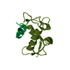
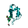
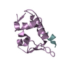
ProteinMdm4 / Double minute 4 protein / Mdm2-like p53-binding protein / Protein Mdmx / p53-binding protein Mdm4
Mass: 9827.411 Da / Num. of mol.: 2 / Source method: obtained synthetically Details: Synthesized based on the human MDMX sequence residues 24-108 and Tyrosine at position 99 is phosphorylated Source: (synth.) Homo sapiens (human) / References: UniProt: O15151
#2: Protein/peptide
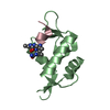
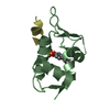
12-MERPEPTIDEINHIBITOR
Mass: 1427.557 Da / Num. of mol.: 2 / Source method: obtained synthetically / Details: 12-mer peptide inhibitor based on human p53
In the structure databanks used in Yorodumi, some data are registered as the other names, "COVID-19 virus" and "2019-nCoV". Here are the details of the virus and the list of structure data.
Jan 31, 2019. EMDB accession codes are about to change! (news from PDBe EMDB page)
EMDB accession codes are about to change! (news from PDBe EMDB page)
The allocation of 4 digits for EMDB accession codes will soon come to an end. Whilst these codes will remain in use, new EMDB accession codes will include an additional digit and will expand incrementally as the available range of codes is exhausted. The current 4-digit format prefixed with “EMD-” (i.e. EMD-XXXX) will advance to a 5-digit format (i.e. EMD-XXXXX), and so on. It is currently estimated that the 4-digit codes will be depleted around Spring 2019, at which point the 5-digit format will come into force.
The EM Navigator/Yorodumi systems omit the EMD- prefix.
Related info.:Q: What is EMD? / ID/Accession-code notation in Yorodumi/EM Navigator
Yorodumi is a browser for structure data from EMDB, PDB, SASBDB, etc.
This page is also the successor to EM Navigator detail page, and also detail information page/front-end page for Omokage search.
The word "yorodu" (or yorozu) is an old Japanese word meaning "ten thousand". "mi" (miru) is to see.
Related info.:EMDB / PDB / SASBDB / Comparison of 3 databanks / Yorodumi Search / Aug 31, 2016. New EM Navigator & Yorodumi / Yorodumi Papers / Jmol/JSmol / Function and homology information / Changes in new EM Navigator and Yorodumi
 Movie
Movie Controller

 Yorodumi
 Open data
 Basic information
 Components
 Keywords
 Function and homology information
 Homo sapiens (human)
 X-RAY DIFFRACTION /
  Authors
 Citation
 Structure visualization
 Downloads & links
 Other downloads


 PDBj
PDBj

 Assembly
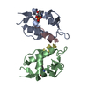

 Mass: 18.015 Da / Num. of mol.: 141 / Source method: isolated from a natural source / Formula: H2O
 Sample preparation
 / Beamline: BL7-1 / Wavelength: 0.9753 Å
 Processing