 Movie
Movie Controller

[English] 日本語
 Yorodumi
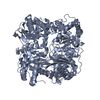
- PDB-4pes: Crystal structure of insulin degrading enzyme complexed with inhi... -
+ Open data

- Basic information
| Entry | Database: PDB / ID: 4pes | |||||||||
|---|---|---|---|---|---|---|---|---|---|---|
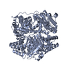
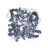
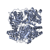
| Title | Crystal structure of insulin degrading enzyme complexed with inhibitor tert-butyl [(2S)-2-(2,5-difluorophenyl)-3-(quinolin-3-yl)propyl]carbamate | |||||||||
 Components Components |
| |||||||||
 Keywords Keywords | HYDROLASE/HYDROLASE INHIBITOR / HYDROLASE-HYDROLASE INHIBITOR complex | |||||||||
| Function / homology |  Function and homology information Function and homology informationinsulysin / beta-endorphin binding / ubiquitin recycling / insulin catabolic process / insulin metabolic process / amyloid-beta clearance by cellular catabolic process / hormone catabolic process / bradykinin catabolic process / cytosolic proteasome complex / positive regulation of protein binding ...insulysin / beta-endorphin binding / ubiquitin recycling / insulin catabolic process / insulin metabolic process / amyloid-beta clearance by cellular catabolic process / hormone catabolic process / bradykinin catabolic process / cytosolic proteasome complex / positive regulation of protein binding / insulin binding / regulation of aerobic respiration / peptide catabolic process / peroxisomal matrix / amyloid-beta clearance / amyloid-beta metabolic process / Insulin receptor recycling / negative regulation of proteolysis / peptide binding / : / protein catabolic process / Peroxisomal protein import / antigen processing and presentation of endogenous peptide antigen via MHC class I / metalloendopeptidase activity / positive regulation of protein catabolic process / insulin receptor signaling pathway / peroxisome / amyloid-beta binding / virus receptor activity / endopeptidase activity / basolateral plasma membrane / Ub-specific processing proteases / external side of plasma membrane / protein-containing complex binding / cell surface / protein homodimerization activity / ATP hydrolysis activity / mitochondrion / proteolysis / : / extracellular exosome / zinc ion binding / ATP binding / identical protein binding / nucleus / cytosol / cytoplasm Similarity search - Function | |||||||||
| Biological species |  Homo sapiens (human) Homo sapiens (human) | |||||||||
| Method |  X-RAY DIFFRACTION / SYNCHROTRON / MOLECULAR REPLACEMENT / Resolution: 2.21 Å X-RAY DIFFRACTION / SYNCHROTRON / MOLECULAR REPLACEMENT / Resolution: 2.21 Å | |||||||||
 Authors Authors | Wang, Y. / Guo, S. | |||||||||
 Citation Citation | Journal: To Be Published Title: Crystal structure of IDE complexed with an inhibitor Authors: Durham, T.B. / Toth, J.L. / Klimkowski, V.J. / Cao, J.X.C. / Siesky, A.M. / Alexander-Chacko, J. / Wu, G.Y. / Dixon, J.T. / McGee, J.E. / Wang, Y. / Guo, S. / Cavitt, R.N. / Schindler, J. / ...Authors: Durham, T.B. / Toth, J.L. / Klimkowski, V.J. / Cao, J.X.C. / Siesky, A.M. / Alexander-Chacko, J. / Wu, G.Y. / Dixon, J.T. / McGee, J.E. / Wang, Y. / Guo, S. / Cavitt, R.N. / Schindler, J. / Thibodeaux, S.J. / Calvert, N.A. / Coghlan, M.J. / Sindelar, D.K. / Christe, M. / Kiselyov, V.V. / Dodson, M. / Sloop, K.W. | |||||||||
| History |
|
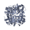
- Structure visualization
| Structure viewer | Molecule: MolmilJmol/JSmol |
|---|
- Downloads & links
-Download
| PDBx/mmCIF format | 4pes.cif.gz | 414.1 KB | Display | PDBx/mmCIF format |
|---|---|---|---|---|
| PDB format | pdb4pes.ent.gz | 327.5 KB | Display | PDB format |
| PDBx/mmJSON format | 4pes.json.gz | Tree view | PDBx/mmJSON format | |
| Others |  Other downloads Other downloads |
-Validation report
| Arichive directory | https://data.pdbj.org/pub/pdb/validation_reports/pe/4pesftp://data.pdbj.org/pub/pdb/validation_reports/pe/4pes | HTTPS FTP |
|---|
-Related structure data
| Related structure data |  2g47S S: Starting model for refinement |
|---|---|
| Similar structure data |


-Links
 PDBj
PDBj


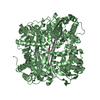
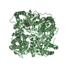
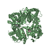
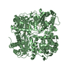
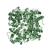
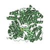
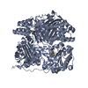
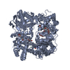
- Assembly
| Deposited unit | 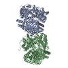
| ||||||||||||||||||
|---|---|---|---|---|---|---|---|---|---|---|---|---|---|---|---|---|---|---|---|
| 1 | 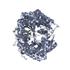
| ||||||||||||||||||
| 2 | 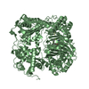
| ||||||||||||||||||
| Unit cell |
| ||||||||||||||||||
| Noncrystallographic symmetry (NCS) | NCS domain:
NCS domain segments: Component-ID: _ / Ens-ID: 1 / Beg auth comp-ID: ASN / Beg label comp-ID: ASN / End auth comp-ID: ILE / End label comp-ID: ILE / Refine code: _ / Auth seq-ID: 44 - 1012 / Label seq-ID: 14 - 982
|
-Components
| #1: Protein | Mass: 114493.484 Da / Num. of mol.: 2 / Fragment: UNP residues 42-1019 Mutation: C110L, E111Q, C171S, C178A, C257V, C414L, C573N, C590S, C789S, C812A, C819A, C904S, C966N, C974A Source method: isolated from a genetically manipulated source Source: (gene. exp.) Homo sapiens (human) / Gene: IDE / Production host:  #2: Protein/peptide | Mass: 231.249 Da / Num. of mol.: 2 Source method: isolated from a genetically manipulated source Source: (gene. exp.) Homo sapiens (human) / Production host: #3: Chemical | 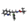  Mass: 398.446 Da / Num. of mol.: 2 / Source method: obtained synthetically / Formula: C23H24F2N2O2 Mass: 398.446 Da / Num. of mol.: 2 / Source method: obtained synthetically / Formula: C23H24F2N2O2#4: Chemical |   Mass: 65.409 Da / Num. of mol.: 2 / Source method: obtained synthetically / Formula: Zn Mass: 65.409 Da / Num. of mol.: 2 / Source method: obtained synthetically / Formula: Zn#5: Water | ChemComp-HOH / |  Mass: 18.015 Da / Num. of mol.: 472 / Source method: isolated from a natural source / Formula: H2O Mass: 18.015 Da / Num. of mol.: 472 / Source method: isolated from a natural source / Formula: H2O |
|---|
-Experimental details
-Experiment
| Experiment | Method: X-RAY DIFFRACTION |
|---|
- Sample preparation
| Crystal | Density Matthews: 2.44 Å3/Da / Density % sol: 49.68 % |
|---|---|
| Crystal grow | Temperature: 298 K / Method: vapor diffusion / Details: 20% PEG3350, 0.2 mM sodium thiocyanate |
-Data collection
| Diffraction | Mean temperature: 100 K |
|---|---|
| Diffraction source | Source: SYNCHROTRON / Site: APS  / Beamline: 31-ID / Wavelength: 0.97931 Å / Beamline: 31-ID / Wavelength: 0.97931 Å |
| Detector | Type: RAYONIX MX225HE / Detector: CCD / Date: Oct 12, 2011 |
| Radiation | Monochromator: diamond(111) / Protocol: SINGLE WAVELENGTH / Monochromatic (M) / Laue (L): M / Scattering type: x-ray |
| Radiation wavelength | Wavelength: 0.97931 Å / Relative weight: 1 |
| Reflection | Resolution: 2.2→122.69 Å / Num. obs: 119894 / % possible obs: 98.8 % / Redundancy: 3.7 % / Rsym value: 0.07 / Net I/σ(I): 7.9 |
| Reflection shell | Highest resolution: 2.2 Å / Rsym value: 0.606 |
- Processing
| Software |
| ||||||||||||||||||||||||||||||||||||||||||||||||||||||||||||||||||||||||||||||||||||||||||||||||||||||||||||||||||||||||||||||||||||||||||||||||||||||||||||||||||||||||||||||||||||||
|---|---|---|---|---|---|---|---|---|---|---|---|---|---|---|---|---|---|---|---|---|---|---|---|---|---|---|---|---|---|---|---|---|---|---|---|---|---|---|---|---|---|---|---|---|---|---|---|---|---|---|---|---|---|---|---|---|---|---|---|---|---|---|---|---|---|---|---|---|---|---|---|---|---|---|---|---|---|---|---|---|---|---|---|---|---|---|---|---|---|---|---|---|---|---|---|---|---|---|---|---|---|---|---|---|---|---|---|---|---|---|---|---|---|---|---|---|---|---|---|---|---|---|---|---|---|---|---|---|---|---|---|---|---|---|---|---|---|---|---|---|---|---|---|---|---|---|---|---|---|---|---|---|---|---|---|---|---|---|---|---|---|---|---|---|---|---|---|---|---|---|---|---|---|---|---|---|---|---|---|---|---|---|---|
| Refinement | Method to determine structure: MOLECULAR REPLACEMENT Starting model: PDB entry 2G47 Resolution: 2.21→122.69 Å / Cor.coef. Fo:Fc: 0.953 / Cor.coef. Fo:Fc free: 0.93 / SU B: 8.065 / SU ML: 0.194 / Cross valid method: THROUGHOUT / ESU R: 0.285 / ESU R Free: 0.221 / Stereochemistry target values: MAXIMUM LIKELIHOOD
| ||||||||||||||||||||||||||||||||||||||||||||||||||||||||||||||||||||||||||||||||||||||||||||||||||||||||||||||||||||||||||||||||||||||||||||||||||||||||||||||||||||||||||||||||||||||
| Solvent computation | Ion probe radii: 0.8 Å / Shrinkage radii: 0.8 Å / VDW probe radii: 1.2 Å / Solvent model: BABINET MODEL WITH MASK | ||||||||||||||||||||||||||||||||||||||||||||||||||||||||||||||||||||||||||||||||||||||||||||||||||||||||||||||||||||||||||||||||||||||||||||||||||||||||||||||||||||||||||||||||||||||
| Displacement parameters | Biso mean: 46.756 Å2
| ||||||||||||||||||||||||||||||||||||||||||||||||||||||||||||||||||||||||||||||||||||||||||||||||||||||||||||||||||||||||||||||||||||||||||||||||||||||||||||||||||||||||||||||||||||||
| Refinement step | Cycle: 1 / Resolution: 2.21→122.69 Å
| ||||||||||||||||||||||||||||||||||||||||||||||||||||||||||||||||||||||||||||||||||||||||||||||||||||||||||||||||||||||||||||||||||||||||||||||||||||||||||||||||||||||||||||||||||||||
| Refine LS restraints |
|