 Movie
Movie Controller


+ Open data

- Basic information
| Entry | Database: PDB / ID: 4pdo | ||||||
|---|---|---|---|---|---|---|---|
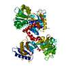
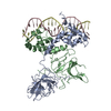
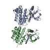
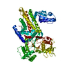
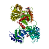
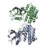
| Title | Structure of Ephrin type-A receptor 2 | ||||||
 Components Components | Ephrin type-A receptor 2 | ||||||
 Keywords Keywords | TRANSFERASE / EphA2 | ||||||
| Function / homology |  Function and homology information Function and homology informationnotochord cell development / notochord formation / lens fiber cell morphogenesis / blood vessel endothelial cell proliferation involved in sprouting angiogenesis / negative regulation of lymphangiogenesis / axial mesoderm formation / cAMP metabolic process / regulation of blood vessel endothelial cell migration / pericyte cell differentiation / leading edge membrane ...notochord cell development / notochord formation / lens fiber cell morphogenesis / blood vessel endothelial cell proliferation involved in sprouting angiogenesis / negative regulation of lymphangiogenesis / axial mesoderm formation / cAMP metabolic process / regulation of blood vessel endothelial cell migration / pericyte cell differentiation / leading edge membrane / negative regulation of chemokine production / ephrin receptor activity / activation of GTPase activity / response to growth factor / post-anal tail morphogenesis / bone remodeling / positive regulation of bicellular tight junction assembly / regulation of lamellipodium assembly / negative regulation of cell adhesion mediated by integrin / branching involved in mammary gland duct morphogenesis / central nervous system neuron differentiation / EPH-Ephrin signaling / neural tube development / RND1 GTPase cycle / RND2 GTPase cycle / RND3 GTPase cycle / mammary gland epithelial cell proliferation / tight junction / RHOV GTPase cycle / EPHA-mediated growth cone collapse / growth factor binding / RHOU GTPase cycle / lamellipodium membrane / RHOG GTPase cycle / EPH-ephrin mediated repulsion of cells / RAC3 GTPase cycle / regulation of angiogenesis / RAC2 GTPase cycle / ephrin receptor signaling pathway / vasculogenesis / regulation of ERK1 and ERK2 cascade / keratinocyte differentiation / RAC1 GTPase cycle / transmembrane receptor protein tyrosine kinase activity / osteoclast differentiation / negative regulation of angiogenesis / cell surface receptor protein tyrosine kinase signaling pathway / molecular function activator activity / protein localization to plasma membrane / positive regulation of protein localization to plasma membrane / cell chemotaxis / skeletal system development / cell motility / receptor protein-tyrosine kinase / intrinsic apoptotic signaling pathway in response to DNA damage / ruffle membrane / osteoblast differentiation / cell migration / lamellipodium / virus receptor activity / angiogenesis / cell adhesion / signaling receptor complex / defense response to Gram-positive bacterium / positive regulation of cell migration / cadherin binding / inflammatory response / focal adhesion / cell surface / ATP binding / plasma membrane Similarity search - Function | ||||||
| Biological species |  Homo sapiens (human) Homo sapiens (human) | ||||||
| Method |  X-RAY DIFFRACTION / SYNCHROTRON / MOLECULAR REPLACEMENT / Resolution: 2.104 Å X-RAY DIFFRACTION / SYNCHROTRON / MOLECULAR REPLACEMENT / Resolution: 2.104 Å | ||||||
 Authors Authors | Jimin, Z. / Qiang, W. / Zongchao, J. | ||||||
 Citation Citation | Journal: To Be Published Title: Structure of Ephrin type-A receptor 2 Authors: Nan, W. | ||||||
| History |
|
- Structure visualization
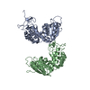
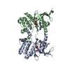
| Structure viewer | Molecule: MolmilJmol/JSmol |
|---|
- Downloads & links
-Download
| PDBx/mmCIF format | 4pdo.cif.gz | 128.6 KB | Display | PDBx/mmCIF format |
|---|---|---|---|---|
| PDB format | pdb4pdo.ent.gz | 97.9 KB | Display | PDB format |
| PDBx/mmJSON format | 4pdo.json.gz | Tree view | PDBx/mmJSON format | |
| Others |  Other downloads Other downloads |
-Validation report
| Arichive directory | https://data.pdbj.org/pub/pdb/validation_reports/pd/4pdoftp://data.pdbj.org/pub/pdb/validation_reports/pd/4pdo | HTTPS FTP |
|---|
-Related structure data
| Related structure data |  4p2kC  4trlC 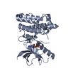 1mqbS C: citing same article ( S: Starting model for refinement |
|---|---|
| Similar structure data |


-Links
 PDBj
PDBj


- Assembly
| Deposited unit | 
| ||||||||
|---|---|---|---|---|---|---|---|---|---|
| 1 |
| ||||||||
| Unit cell |
| ||||||||
| Details | biological unit is the same as asym. |
-Components
| #1: Protein | Mass: 34027.176 Da / Num. of mol.: 2 / Fragment: UNP residues 590-876 Source method: isolated from a genetically manipulated source Source: (gene. exp.) Homo sapiens (human) / Gene: EPHA2, ECK / Production host:  References: UniProt: P29317, receptor protein-tyrosine kinase #2: Water | ChemComp-HOH / |  Mass: 18.015 Da / Num. of mol.: 481 / Source method: isolated from a natural source / Formula: H2O Mass: 18.015 Da / Num. of mol.: 481 / Source method: isolated from a natural source / Formula: H2O |
|---|
-Experimental details
-Experiment
| Experiment | Method: X-RAY DIFFRACTION / Number of used crystals: 1 |
|---|
- Sample preparation
| Crystal | Density Matthews: 2.75 Å3/Da / Density % sol: 55.27 % |
|---|---|
| Crystal grow | Temperature: 293 K / Method: vapor diffusion, hanging drop / pH: 8.5 Details: 0.8 M Na/K L(+)-tartrate, 0.1 M Tris-Cl pH 8.5, 1% (w/v) PEGMME 5000 |
-Data collection
| Diffraction | Mean temperature: 100 K | ||||||||||||||||||||||||||||||||||||||||||||||||||||||||||||||||||||||||||||||||||||||||||||||||||||||||||||||||||||||||||||||
|---|---|---|---|---|---|---|---|---|---|---|---|---|---|---|---|---|---|---|---|---|---|---|---|---|---|---|---|---|---|---|---|---|---|---|---|---|---|---|---|---|---|---|---|---|---|---|---|---|---|---|---|---|---|---|---|---|---|---|---|---|---|---|---|---|---|---|---|---|---|---|---|---|---|---|---|---|---|---|---|---|---|---|---|---|---|---|---|---|---|---|---|---|---|---|---|---|---|---|---|---|---|---|---|---|---|---|---|---|---|---|---|---|---|---|---|---|---|---|---|---|---|---|---|---|---|---|---|
| Diffraction source | Source: SYNCHROTRON / Site: Photon Factory  / Beamline: BL-5A / Wavelength: 1 Å / Beamline: BL-5A / Wavelength: 1 Å | ||||||||||||||||||||||||||||||||||||||||||||||||||||||||||||||||||||||||||||||||||||||||||||||||||||||||||||||||||||||||||||||
| Detector | Type: ADSC QUANTUM 315r / Detector: CCD / Date: Nov 23, 2013 | ||||||||||||||||||||||||||||||||||||||||||||||||||||||||||||||||||||||||||||||||||||||||||||||||||||||||||||||||||||||||||||||
| Radiation | Protocol: SINGLE WAVELENGTH / Monochromatic (M) / Laue (L): M / Scattering type: x-ray | ||||||||||||||||||||||||||||||||||||||||||||||||||||||||||||||||||||||||||||||||||||||||||||||||||||||||||||||||||||||||||||||
| Radiation wavelength | Wavelength: 1 Å / Relative weight: 1 | ||||||||||||||||||||||||||||||||||||||||||||||||||||||||||||||||||||||||||||||||||||||||||||||||||||||||||||||||||||||||||||||
| Reflection | Resolution: 2.1→15 Å / Num. obs: 41909 / % possible obs: 99.8 % / Redundancy: 4.7 % / Rmerge(I) obs: 0.086 / Χ2: 0.902 / Net I/av σ(I): 19.445 / Net I/σ(I): 9.3 / Num. measured all: 197356 | ||||||||||||||||||||||||||||||||||||||||||||||||||||||||||||||||||||||||||||||||||||||||||||||||||||||||||||||||||||||||||||||
| Reflection shell | Diffraction-ID: 1 / Rejects: _
|
- Processing
| Software |
| ||||||||||||||||||||||||||||||||||||||||||||||||||||||||||||||||||||||||||||||||||||||||||||||||||||||||||||||||
|---|---|---|---|---|---|---|---|---|---|---|---|---|---|---|---|---|---|---|---|---|---|---|---|---|---|---|---|---|---|---|---|---|---|---|---|---|---|---|---|---|---|---|---|---|---|---|---|---|---|---|---|---|---|---|---|---|---|---|---|---|---|---|---|---|---|---|---|---|---|---|---|---|---|---|---|---|---|---|---|---|---|---|---|---|---|---|---|---|---|---|---|---|---|---|---|---|---|---|---|---|---|---|---|---|---|---|---|---|---|---|---|---|---|
| Refinement | Method to determine structure: MOLECULAR REPLACEMENT Starting model: 1MQB Resolution: 2.104→14.902 Å / FOM work R set: 0.9004 / SU ML: 0.18 / Cross valid method: FREE R-VALUE / σ(F): 1.99 / Phase error: 17.31 / Stereochemistry target values: ML
| ||||||||||||||||||||||||||||||||||||||||||||||||||||||||||||||||||||||||||||||||||||||||||||||||||||||||||||||||
| Solvent computation | Shrinkage radii: 0.9 Å / VDW probe radii: 1.11 Å / Solvent model: FLAT BULK SOLVENT MODEL | ||||||||||||||||||||||||||||||||||||||||||||||||||||||||||||||||||||||||||||||||||||||||||||||||||||||||||||||||
| Displacement parameters | Biso max: 58.72 Å2 / Biso mean: 20.64 Å2 / Biso min: 6.71 Å2 | ||||||||||||||||||||||||||||||||||||||||||||||||||||||||||||||||||||||||||||||||||||||||||||||||||||||||||||||||
| Refinement step | Cycle: final / Resolution: 2.104→14.902 Å
| ||||||||||||||||||||||||||||||||||||||||||||||||||||||||||||||||||||||||||||||||||||||||||||||||||||||||||||||||
| Refine LS restraints |
| ||||||||||||||||||||||||||||||||||||||||||||||||||||||||||||||||||||||||||||||||||||||||||||||||||||||||||||||||
| LS refinement shell | Refine-ID: X-RAY DIFFRACTION / Total num. of bins used: 15
|
