Mass: 18.015 Da / Num. of mol.: 1133 / Source method: isolated from a natural source / Formula: H2O
Has protein modification
Y
-
Experimental details
-
Experiment
Experiment
Method: X-RAY DIFFRACTION
-
Sample preparation
Crystal
Density Matthews: 2.26 Å3/Da / Density % sol: 45.53 %
Crystal grow
Temperature: 295 K / Method: vapor diffusion, hanging drop / pH: 4.8 Details: 0.1 M phosphate citrate buffer, 2 M ammonium sulphate PH range: 4.2-4.8 / Temp details: 295
-
Data collection
Diffraction
Mean temperature: 100 K
Diffraction source
Source: ROTATING ANODE / Type: BRUKER AXS MICROSTAR / Wavelength: 1.5418 Å
Detector
Type: MAR scanner 345 mm plate / Detector: IMAGE PLATE / Date: Nov 10, 2014
Radiation
Protocol: SINGLE WAVELENGTH / Monochromatic (M) / Laue (L): M / Scattering type: x-ray
Radiation wavelength
Wavelength: 1.5418 Å / Relative weight: 1
Reflection
Resolution: 1.8→20 Å / Num. obs: 76051 / % possible obs: 99.9 % / Redundancy: 10.1 % / Net I/σ(I): 19.97
Reflection shell
Resolution: 1.8→1.9 Å / Redundancy: 10 % / Mean I/σ(I) obs: 3.66 / % possible all: 99.9
-
Processing
Software
Name
Version
Classification
REFMAC
5.7.0032
refinement
XDS
datareduction
XSCALE
datascaling
REFMAC
phasing
Refinement
Resolution: 1.8→19.81 Å / Cor.coef. Fo:Fc: 0.954 / Cor.coef. Fo:Fc free: 0.924 / SU B: 6.415 / SU ML: 0.108 / Cross valid method: THROUGHOUT / ESU R: 0.132 / ESU R Free: 0.133 / Stereochemistry target values: MAXIMUM LIKELIHOOD / Details: HYDROGENS HAVE BEEN ADDED IN THE RIDING POSITIONS
Rfactor
Num. reflection
% reflection
Selection details
Rfree
0.22936
3803
5 %
RANDOM
Rwork
0.17721
-
-
-
obs
0.1798
72247
99.86 %
-
Solvent computation
Ion probe radii: 0.8 Å / Shrinkage radii: 0.8 Å / VDW probe radii: 1.2 Å / Solvent model: MASK
Displacement parameters
Biso mean: 22.599 Å2
Baniso -1
Baniso -2
Baniso -3
1-
2.31 Å2
0 Å2
-0 Å2
2-
-
-0.74 Å2
-0 Å2
3-
-
-
-1.57 Å2
Refinement step
Cycle: 1 / Resolution: 1.8→19.81 Å
Protein
Nucleic acid
Ligand
Solvent
Total
Num. atoms
6002
0
46
1138
7186
Refine LS restraints
Refine-ID
Type
Dev ideal
Dev ideal target
Number
X-RAY DIFFRACTION
r_bond_refined_d
0.019
0.019
6206
X-RAY DIFFRACTION
r_bond_other_d
0.002
0.02
5829
X-RAY DIFFRACTION
r_angle_refined_deg
1.902
1.941
8460
X-RAY DIFFRACTION
r_angle_other_deg
0.931
3.001
13394
X-RAY DIFFRACTION
r_dihedral_angle_1_deg
6.408
5
802
X-RAY DIFFRACTION
r_dihedral_angle_2_deg
36.04
25.889
270
X-RAY DIFFRACTION
r_dihedral_angle_3_deg
14.504
15
965
X-RAY DIFFRACTION
r_dihedral_angle_4_deg
17.909
15
15
X-RAY DIFFRACTION
r_chiral_restr
0.118
0.2
935
X-RAY DIFFRACTION
r_gen_planes_refined
0.01
0.021
7202
X-RAY DIFFRACTION
r_gen_planes_other
0.001
0.02
1389
X-RAY DIFFRACTION
r_mcbond_it
0.652
0.784
3181
X-RAY DIFFRACTION
r_mcbond_other
0.648
0.784
3180
X-RAY DIFFRACTION
r_mcangle_it
1.046
1.173
3977
X-RAY DIFFRACTION
r_mcangle_other
1.046
1.174
3978
X-RAY DIFFRACTION
r_scbond_it
1.004
0.884
3025
X-RAY DIFFRACTION
r_scbond_other
0.99
0.878
3017
X-RAY DIFFRACTION
r_scangle_other
1.546
1.277
4467
X-RAY DIFFRACTION
r_long_range_B_refined
6.433
9.005
8406
X-RAY DIFFRACTION
r_long_range_B_other
6.17
7.243
7708
LS refinement shell
Resolution: 1.8→1.846 Å / Total num. of bins used: 20
Rfactor
Num. reflection
% reflection
Rfree
0.314
274
-
Rwork
0.267
5208
-
obs
-
-
99.98 %
Refinement TLS params.
Method: refined / Refine-ID: X-RAY DIFFRACTION
ID
L11 (°2)
L12 (°2)
L13 (°2)
L22 (°2)
L23 (°2)
L33 (°2)
S11 (Å °)
S12 (Å °)
S13 (Å °)
S21 (Å °)
S22 (Å °)
S23 (Å °)
S31 (Å °)
S32 (Å °)
S33 (Å °)
T11 (Å2)
T12 (Å2)
T13 (Å2)
T22 (Å2)
T23 (Å2)
T33 (Å2)
Origin x (Å)
Origin y (Å)
Origin z (Å)
1
1.635
0.7346
-1.2529
3.2867
-2.3216
2.9079
-0.106
0.28
-0.0645
-0.3593
0.1649
0.1143
0.1181
-0.4158
-0.0589
0.0682
-0.008
-0.0088
0.1553
-0.0287
0.0629
68.574
19.894
45.381
2
0.5565
0.2565
0.2619
1.4352
0.0531
2.7866
0.0512
-0.0461
-0.0755
0.1108
-0.0318
-0.174
-0.028
-0.0269
-0.0194
0.0127
-0.0074
-0.0129
0.0099
-0.0048
0.0644
79.882
14.298
60.885
3
3.8152
-0.0641
1.2245
10.1972
3.7508
3.38
0.2375
0.2413
-1.0006
0.9879
0.0211
-0.7755
0.5175
0.4187
-0.2586
0.2427
-0.0135
-0.1814
0.1216
-0.0165
0.5388
78.267
-9.154
65.92
4
0.6475
0.1741
-0.5604
0.617
-0.1803
2.6761
0.0342
-0.0451
0.0308
-0.0645
-0.0419
-0.0192
-0.2431
-0.1894
0.0077
0.0426
0.0303
-0.0235
0.0325
-0.0195
0.0697
62.186
-15.557
42.83
5
5.0446
-3.5372
-3.004
2.5112
1.6346
9.5792
-0.3173
-0.1012
0.5935
0.3724
0.123
-0.4496
-3.0211
-1.4806
0.1943
1.3176
0.6766
-0.2941
0.5585
-0.1214
0.1665
53.92
-7.492
28.062
6
1.3194
0.3784
-1.2257
0.8928
-0.6513
3.696
-0.0336
0.1176
-0.0689
-0.1346
0.0049
-0.0349
-0.1139
-0.3932
0.0287
0.0456
0.0367
-0.0092
0.0616
-0.021
0.0334
61.78
-18.595
41.963
Refinement TLS group
ID
Refine-ID
Refine TLS-ID
Auth asym-ID
Auth seq-ID
1
X-RAY DIFFRACTION
1
A
26 - 84
2
X-RAY DIFFRACTION
2
A
85 - 402
3
X-RAY DIFFRACTION
3
A
403 - 421
4
X-RAY DIFFRACTION
4
B
26 - 238
5
X-RAY DIFFRACTION
5
B
239 - 266
6
X-RAY DIFFRACTION
6
B
267 - 421
+
About Yorodumi
-
News
-
Feb 9, 2022. New format data for meta-information of EMDB entries
New format data for meta-information of EMDB entries
Version 3 of the EMDB header file is now the official format.
The previous official version 1.9 will be removed from the archive.
In the structure databanks used in Yorodumi, some data are registered as the other names, "COVID-19 virus" and "2019-nCoV". Here are the details of the virus and the list of structure data.
Jan 31, 2019. EMDB accession codes are about to change! (news from PDBe EMDB page)
EMDB accession codes are about to change! (news from PDBe EMDB page)
The allocation of 4 digits for EMDB accession codes will soon come to an end. Whilst these codes will remain in use, new EMDB accession codes will include an additional digit and will expand incrementally as the available range of codes is exhausted. The current 4-digit format prefixed with “EMD-” (i.e. EMD-XXXX) will advance to a 5-digit format (i.e. EMD-XXXXX), and so on. It is currently estimated that the 4-digit codes will be depleted around Spring 2019, at which point the 5-digit format will come into force.
The EM Navigator/Yorodumi systems omit the EMD- prefix.
Related info.:Q: What is EMD? / ID/Accession-code notation in Yorodumi/EM Navigator
Yorodumi is a browser for structure data from EMDB, PDB, SASBDB, etc.
This page is also the successor to EM Navigator detail page, and also detail information page/front-end page for Omokage search.
The word "yorodu" (or yorozu) is an old Japanese word meaning "ten thousand". "mi" (miru) is to see.
Related info.:EMDB / PDB / SASBDB / Comparison of 3 databanks / Yorodumi Search / Aug 31, 2016. New EM Navigator & Yorodumi / Yorodumi Papers / Jmol/JSmol / Function and homology information / Changes in new EM Navigator and Yorodumi
 Movie
Movie Controller

 Yorodumi
 Open data
 Basic information
 Components
 Keywords
 Function and homology information
 Pseudomonas putida CSV86 (bacteria)
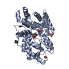
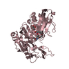
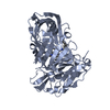
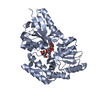
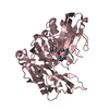
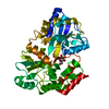
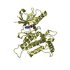
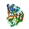
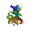
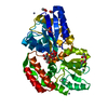
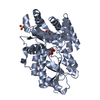
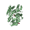
 X-RAY DIFFRACTION / Resolution: 1.8 Å
  Authors
 India, 2items
  Citation
 Structure visualization
 Downloads & links
 Other downloads

 PDBj
PDBj


 Assembly

 Type: D-saccharide, beta linking / Mass: 180.156 Da / Num. of mol.: 2
 Mass: 92.094 Da / Num. of mol.: 2 / Source method: obtained synthetically / Formula: C3H8O3
 Mass: 96.063 Da / Num. of mol.: 2 / Source method: obtained synthetically / Formula: SO4
 Mass: 18.015 Da / Num. of mol.: 1133 / Source method: isolated from a natural source / Formula: H2O
 Sample preparation
 Processing