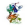
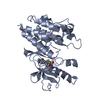
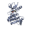
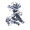
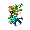
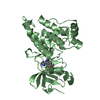
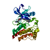
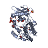
[English] 日本語
Yorodumi
- PDB-4trl: Structure of Ephrin type-A receptor 2 -

+
Open data


ID or keywords:

Loading...

-
Basic information

Entry
Database: PDB / ID: 4trl
TitleStructure of Ephrin type-A receptor 2
ComponentsEphrin type-A receptor 2
KeywordsTRANSFERASE / active state / phosphorylated
Function / homology
Function and homology information


notochord cell development / notochord formation / lens fiber cell morphogenesis / blood vessel endothelial cell proliferation involved in sprouting angiogenesis / negative regulation of lymphangiogenesis / axial mesoderm formation / pericyte cell differentiation / cAMP metabolic process / positive regulation of bicellular tight junction assembly / regulation of blood vessel endothelial cell migration ...notochord cell development / notochord formation / lens fiber cell morphogenesis / blood vessel endothelial cell proliferation involved in sprouting angiogenesis / negative regulation of lymphangiogenesis / axial mesoderm formation / pericyte cell differentiation / cAMP metabolic process / positive regulation of bicellular tight junction assembly / regulation of blood vessel endothelial cell migration / leading edge membrane / negative regulation of chemokine production / post-anal tail morphogenesis / bone remodeling / transmembrane-ephrin receptor activity / response to growth factor / tight junction / activation of GTPase activity / regulation of lamellipodium assembly / branching involved in mammary gland duct morphogenesis / EPH-Ephrin signaling / neural tube development / RND1 GTPase cycle / RND2 GTPase cycle / RND3 GTPase cycle / mammary gland epithelial cell proliferation / RHOV GTPase cycle / EPHA-mediated growth cone collapse / growth factor binding / regulation of cell adhesion mediated by integrin / lamellipodium membrane / RHOU GTPase cycle / RHOG GTPase cycle / EPH-ephrin mediated repulsion of cells / ephrin receptor signaling pathway / RAC2 GTPase cycle / RAC3 GTPase cycle / negative regulation of phosphatidylinositol 3-kinase/protein kinase B signal transduction / vasculogenesis / regulation of angiogenesis / keratinocyte differentiation / RAC1 GTPase cycle / transmembrane receptor protein tyrosine kinase activity / negative regulation of angiogenesis / phosphatidylinositol 3-kinase/protein kinase B signal transduction / regulation of ERK1 and ERK2 cascade / osteoclast differentiation / cell chemotaxis / skeletal system development / molecular function activator activity / protein localization to plasma membrane / cell motility / positive regulation of protein localization to plasma membrane / axon guidance / receptor protein-tyrosine kinase / ruffle membrane / osteoblast differentiation / intrinsic apoptotic signaling pathway in response to DNA damage / cell migration / lamellipodium / virus receptor activity / receptor complex / cell adhesion / defense response to Gram-positive bacterium / positive regulation of cell migration / cadherin binding / inflammatory response / phosphorylation / focal adhesion / dendrite / cell surface / ATP binding / plasma membrane
Similarity search - Function
Ephrin type-A receptor 2, ligand binding domain / Ephrin receptor type-A /type-B / Ephrin receptor ligand binding domain / Tyrosine-protein kinase, receptor class V, conserved site / Ephrin receptor, transmembrane domain / Ephrin receptor ligand binding domain / Ephrin type-A receptor 2 transmembrane domain / Receptor tyrosine kinase class V signature 1. / Receptor tyrosine kinase class V signature 2. / Eph receptor ligand-binding domain profile. ...Ephrin type-A receptor 2, ligand binding domain / Ephrin receptor type-A /type-B / Ephrin receptor ligand binding domain / Tyrosine-protein kinase, receptor class V, conserved site / Ephrin receptor, transmembrane domain / Ephrin receptor ligand binding domain / Ephrin type-A receptor 2 transmembrane domain / Receptor tyrosine kinase class V signature 1. / Receptor tyrosine kinase class V signature 2. / Eph receptor ligand-binding domain profile. / Ephrin receptor ligand binding domain / Putative ephrin-receptor like / SAM domain (Sterile alpha motif) / SAM domain profile. / Sterile alpha motif. / Sterile alpha motif domain / Sterile alpha motif/pointed domain superfamily / Growth factor receptor cysteine-rich domain superfamily / Fibronectin type III domain / Fibronectin type 3 domain / Fibronectin type-III domain profile. / Galactose-binding-like domain superfamily / Fibronectin type III / Fibronectin type III superfamily / Tyrosine-protein kinase, catalytic domain / Tyrosine kinase, catalytic domain / Tyrosine protein kinases specific active-site signature. / Tyrosine-protein kinase, active site / Protein tyrosine and serine/threonine kinase / Serine-threonine/tyrosine-protein kinase, catalytic domain / Transferase(Phosphotransferase) domain 1 / Transferase(Phosphotransferase); domain 1 / Phosphorylase Kinase; domain 1 / Phosphorylase Kinase; domain 1 / Protein kinase, ATP binding site / Protein kinases ATP-binding region signature. / Immunoglobulin-like fold / Protein kinase domain profile. / Protein kinase domain / Protein kinase-like domain superfamily / 2-Layer Sandwich / Orthogonal Bundle / Mainly Alpha / Alpha Beta
Similarity search - Domain/homology
Ephrin type-A receptor 2
Similarity search - Component
Biological speciesHomo sapiens (human)
MethodX-RAY DIFFRACTION / SYNCHROTRON / Resolution: 2.452 Å
AuthorsWei, Q. / Wang, N.
CitationJournal: To Be Published
Title: Structure of Ephrin type-A receptor 2
Authors: Wei, Q. / Wang, N.
History
DepositionJun 17, 2014Deposition site: RCSB / Processing site: RCSB
SupersessionJul 2, 2014ID: 4P2W
Revision 1.0Jul 2, 2014Provider: repository / Type: Initial release
Revision 1.1Nov 22, 2017Group: Data collection / Database references ...Data collection / Database references / Derived calculations / Other / Refinement description / Source and taxonomy / Structure summary
Category: citation / diffrn_source ...citation / diffrn_source / entity_src_gen / pdbx_database_status / pdbx_struct_assembly / pdbx_struct_assembly_prop / pdbx_struct_oper_list / software / struct_keywords
Item: _citation.journal_id_CSD / _diffrn_source.pdbx_synchrotron_site ..._citation.journal_id_CSD / _diffrn_source.pdbx_synchrotron_site / _entity_src_gen.pdbx_alt_source_flag / _pdbx_database_status.pdb_format_compatible / _pdbx_struct_assembly.oligomeric_details / _pdbx_struct_assembly_prop.type / _pdbx_struct_assembly_prop.value / _pdbx_struct_oper_list.symmetry_operation / _struct_keywords.text
Revision 1.2Dec 27, 2023Group: Data collection / Database references / Refinement description
Category: chem_comp_atom / chem_comp_bond ...chem_comp_atom / chem_comp_bond / database_2 / refine_hist
Item: _database_2.pdbx_DOI / _database_2.pdbx_database_accession ..._database_2.pdbx_DOI / _database_2.pdbx_database_accession / _refine_hist.pdbx_number_atoms_nucleic_acid / _refine_hist.pdbx_number_atoms_protein

-
Structure visualization

Structure viewerMolecule:
MolmilJmol/JSmol

Downloads & links

-
Assembly

Deposited unit
A: Ephrin type-A receptor 2


Theoretical massNumber of molelcules
Total (without water)34,1071
Polymers34,1071
Non-polymers00
Water77543
1


  • Idetical with deposited unit
  • defined by author&software
TypeNameSymmetry operationNumber
identity operation1_555x,y,z1
Buried area0 Å2
ΔGint0 kcal/mol
Surface area13340 Å2
MethodPISA
Unit cell
Length a, b, c (Å)74.028, 74.028, 101.950
Angle α, β, γ (deg.)90.000, 90.000, 120.000
Int Tables number152
Space group name H-MP3121

-
Components

#1: Protein Ephrin type-A receptor 2 / Epithelial cell kinase / Tyrosine-protein kinase receptor ECK


Mass: 34107.156 Da / Num. of mol.: 1 / Fragment: UNP residues 590-876
Source method: isolated from a genetically manipulated source
Source: (gene. exp.) Homo sapiens (human) / Gene: EPHA2, ECK / Production host: Escherichia coli (E. coli) / Strain (production host): ArcticExpress
References: UniProt: P29317, receptor protein-tyrosine kinase
#2: Water ChemComp-HOH / water


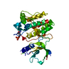
Mass: 18.015 Da / Num. of mol.: 43 / Source method: isolated from a natural source / Formula: H2O

-
Experimental details

-
Experiment

ExperimentMethod: X-RAY DIFFRACTION / Number of used crystals: 1

-
Sample preparation

CrystalDensity Matthews: 2.37 Å3/Da / Density % sol: 48.1 %
Crystal growTemperature: 293 K / Method: vapor diffusion, hanging drop / pH: 7 / Details: 0.1M HEPES, 0.2M Li2SO4, 20% (w/v) PEG3350

-
Data collection

DiffractionMean temperature: 193 K
Diffraction sourceSource: SYNCHROTRON / Site: Photon Factory / Beamline: AR-NW12A / Wavelength: 1 Å
DetectorType: ADSC QUANTUM 210 / Detector: CCD / Date: Nov 6, 2012
RadiationProtocol: SINGLE WAVELENGTH / Monochromatic (M) / Laue (L): M / Scattering type: x-ray
Radiation wavelengthWavelength: 1 Å / Relative weight: 1
ReflectionResolution: 2.45→30 Å / Num. obs: 12263 / % possible obs: 99.8 % / Redundancy: 5.7 % / Biso Wilson estimate: 48.81 Å2 / Rmerge(I) obs: 0.092 / Χ2: 2.212 / Net I/av σ(I): 29.175 / Net I/σ(I): 12.3 / Num. measured all: 69357
Reflection shell

Diffraction-ID: 1 / Rejects: _

Resolution (Å)Redundancy (%)Rmerge(I) obsNum. unique allΧ2% possible all
2.45-2.495.70.6975881.753100
2.49-2.545.80.7036211.699100
2.54-2.595.70.6265801.734100
2.59-2.645.80.5426391.688100
2.64-2.75.70.4095651.733100
2.7-2.765.80.4376171.674100
2.76-2.835.80.3166121.726100
2.83-2.95.80.2485961.731100
2.9-2.995.80.2256081.77100
2.99-3.095.70.1756141.969100
3.09-3.25.90.1436051.952100
3.2-3.325.90.1176062.116100
3.32-3.485.80.0986062.562100
3.48-3.665.80.086172.612100
3.66-3.895.50.0736243.17299.8
3.89-4.195.40.0596112.963100
4.19-4.615.20.0546293.35899.8
4.61-5.275.50.0466292.68799.4
5.27-6.635.60.0526282.61399.4
6.63-3050.0426682.95197.2

-
Processing

Software
NameVersionClassification
SCALEPACKdata scaling
PHENIXrefinement
PDB_EXTRACT3.14data extraction
RefinementResolution: 2.452→30 Å / FOM work R set: 0.7593 / SU ML: 0.36 / Cross valid method: FREE R-VALUE / σ(F): 1.35 / Phase error: 29.51 / Stereochemistry target values: ML
RfactorNum. reflection% reflection
Rfree0.2503 584 4.78 %
Rwork0.199 11642 -
obs0.2015 12226 99.38 %
Solvent computationShrinkage radii: 0.9 Å / VDW probe radii: 1.11 Å / Solvent model: FLAT BULK SOLVENT MODEL
Displacement parametersBiso max: 108.19 Å2 / Biso mean: 38.64 Å2 / Biso min: 15.28 Å2
Refinement stepCycle: final / Resolution: 2.452→30 Å
ProteinNucleic acidLigandSolventTotal
Num. atoms2146 0 0 43 2189
Biso mean---34.91 -
Num. residues----269
Refine LS restraints
Refine-IDTypeDev idealNumber
X-RAY DIFFRACTIONf_bond_d0.0082189
X-RAY DIFFRACTIONf_angle_d1.192949
X-RAY DIFFRACTIONf_chiral_restr0.078324
X-RAY DIFFRACTIONf_plane_restr0.004369
X-RAY DIFFRACTIONf_dihedral_angle_d15.86818
LS refinement shell

Refine-ID: X-RAY DIFFRACTION / Total num. of bins used: 4

Resolution (Å)Rfactor RfreeNum. reflection RfreeRfactor RworkNum. reflection RworkNum. reflection all% reflection obs (%)
2.452-2.69840.381380.27092819295798
2.6984-3.08850.30471630.232828813044100
3.0885-3.88990.25541490.190929073056100
3.8899-30.0280.20831340.18153035316999

+
About Yorodumi

-
News

-
Feb 9, 2022. New format data for meta-information of EMDB entries

New format data for meta-information of EMDB entries

  • Version 3 of the EMDB header file is now the official format.
  • The previous official version 1.9 will be removed from the archive.

Related info.:EMDB header

External links:wwPDB to switch to version 3 of the EMDB data model

-
Aug 12, 2020. Covid-19 info

Covid-19 info

URL: https://pdbjlvh1.pdbj.org/emnavi/covid19.php

New page: Covid-19 featured information page in EM Navigator.

Related info.:Covid-19 info / Mar 5, 2020. Novel coronavirus structure data

+
Mar 5, 2020. Novel coronavirus structure data

Novel coronavirus structure data

Related info.:Yorodumi Speices / Aug 12, 2020. Covid-19 info

External links:COVID-19 featured content - PDBj / Molecule of the Month (242):Coronavirus Proteases

+
Jan 31, 2019. EMDB accession codes are about to change! (news from PDBe EMDB page)

EMDB accession codes are about to change! (news from PDBe EMDB page)

  • The allocation of 4 digits for EMDB accession codes will soon come to an end. Whilst these codes will remain in use, new EMDB accession codes will include an additional digit and will expand incrementally as the available range of codes is exhausted. The current 4-digit format prefixed with “EMD-” (i.e. EMD-XXXX) will advance to a 5-digit format (i.e. EMD-XXXXX), and so on. It is currently estimated that the 4-digit codes will be depleted around Spring 2019, at which point the 5-digit format will come into force.
  • The EM Navigator/Yorodumi systems omit the EMD- prefix.

Related info.:Q: What is EMD? / ID/Accession-code notation in Yorodumi/EM Navigator

External links:EMDB Accession Codes are Changing Soon! / Contact to PDBj

+
Jul 12, 2017. Major update of PDB

Major update of PDB

  • wwPDB released updated PDB data conforming to the new PDBx/mmCIF dictionary.
  • This is a major update changing the version number from 4 to 5, and with Remediation, in which all the entries are updated.
  • In this update, many items about electron microscopy experimental information are reorganized (e.g. em_software).
  • Now, EM Navigator and Yorodumi are based on the updated data.

External links:wwPDB Remediation / Enriched Model Files Conforming to OneDep Data Standards Now Available in the PDB FTP Archive

-
Yorodumi

Thousand views of thousand structures

  • Yorodumi is a browser for structure data from EMDB, PDB, SASBDB, etc.
  • This page is also the successor to EM Navigator detail page, and also detail information page/front-end page for Omokage search.
  • The word "yorodu" (or yorozu) is an old Japanese word meaning "ten thousand". "mi" (miru) is to see.

Related info.:EMDB / PDB / SASBDB / Comparison of 3 databanks / Yorodumi Search / Aug 31, 2016. New EM Navigator & Yorodumi / Yorodumi Papers / Jmol/JSmol / Function and homology information / Changes in new EM Navigator and Yorodumi

Read more