 Movie
Movie Controller

[English] 日本語
 Yorodumi
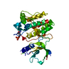
- PDB-1gqz: Refinement of Haemophilus influenzae Diaminopimelate epimerase at 1.7A -
+ Open data

- Basic information
| Entry | Database: PDB / ID: 1gqz | ||||||
|---|---|---|---|---|---|---|---|
| Title | Refinement of Haemophilus influenzae Diaminopimelate epimerase at 1.7A | ||||||
 Components Components | DIAMINOPIMELATE EPIMERASE | ||||||
 Keywords Keywords | ISOMERASE / PEPTIDOGLYCAN BIOSYNTHESIS | ||||||
| Function / homology |  Function and homology information Function and homology informationdiaminopimelate epimerase / diaminopimelate epimerase activity / : / cytosol Similarity search - Function | ||||||
| Biological species |  HAEMOPHILUS INFLUENZAE (bacteria) HAEMOPHILUS INFLUENZAE (bacteria) | ||||||
| Method |  X-RAY DIFFRACTION / SYNCHROTRON / MOLECULAR REPLACEMENT / Resolution: 1.75 Å X-RAY DIFFRACTION / SYNCHROTRON / MOLECULAR REPLACEMENT / Resolution: 1.75 Å | ||||||
 Authors Authors | Roper, D.I. / Huyton, T. / Turkenburg, J.P. | ||||||
 Citation Citation | Journal: Acta Crystallogr. D Biol. Crystallogr. / Year: 2004 Title: Refinement of Haemophilus influenzae diaminopimelic acid epimerase (DapF) at 1.75 A resolution suggests a mechanism for stereocontrol during catalysis. Authors: Lloyd, A.J. / Huyton, T. / Turkenburg, J. / Roper, D.I. | ||||||
| History |
| ||||||
| Remark 700 | SHEET THE SHEET STRUCTURE OF THIS MOLECULE IS BIFURCATED. IN ORDER TO REPRESENT THIS FEATURE IN ... SHEET THE SHEET STRUCTURE OF THIS MOLECULE IS BIFURCATED. IN ORDER TO REPRESENT THIS FEATURE IN THE SHEET RECORDS BELOW, TWO SHEETS ARE DEFINED. |
- Structure visualization
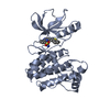
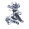
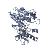
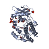
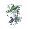
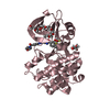
| Structure viewer | Molecule: MolmilJmol/JSmol |
|---|
- Downloads & links
-Download
| PDBx/mmCIF format | 1gqz.cif.gz | 126.9 KB | Display | PDBx/mmCIF format |
|---|---|---|---|---|
| PDB format | pdb1gqz.ent.gz | 99.6 KB | Display | PDB format |
| PDBx/mmJSON format | 1gqz.json.gz | Tree view | PDBx/mmJSON format | |
| Others |  Other downloads Other downloads |
-Validation report
| Arichive directory | https://data.pdbj.org/pub/pdb/validation_reports/gq/1gqzftp://data.pdbj.org/pub/pdb/validation_reports/gq/1gqz | HTTPS FTP |
|---|
-Related structure data
| Related structure data |  1bwzS S: Starting model for refinement |
|---|---|
| Similar structure data |


-Links
 PDBj
PDBj- Assembly
| Deposited unit | 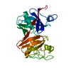
| ||||||||
|---|---|---|---|---|---|---|---|---|---|
| 1 |
| ||||||||
| Unit cell |
|
-Components
| #1: Protein | Mass: 30279.475 Da / Num. of mol.: 1 Source method: isolated from a genetically manipulated source Source: (gene. exp.) HAEMOPHILUS INFLUENZAE (bacteria) / Strain: KW20 / Plasmid: PET28 / Production host: |
|---|---|
| #2: Water | ChemComp-HOH /  Mass: 18.015 Da / Num. of mol.: 353 / Source method: isolated from a natural source / Formula: H2O Mass: 18.015 Da / Num. of mol.: 353 / Source method: isolated from a natural source / Formula: H2O |
| Has protein modification | Y |
-Experimental details
-Experiment
| Experiment | Method: X-RAY DIFFRACTION / Number of used crystals: 1 |
|---|
- Sample preparation
| Crystal | Density Matthews: 2.64 Å3/Da / Density % sol: 53.12 % | ||||||||||||||||||||||||||||||||||||
|---|---|---|---|---|---|---|---|---|---|---|---|---|---|---|---|---|---|---|---|---|---|---|---|---|---|---|---|---|---|---|---|---|---|---|---|---|---|
| Crystal grow | pH: 6.5 / Details: 0.1M IMIDIZOLE PH 6.5, | ||||||||||||||||||||||||||||||||||||
| Crystal grow | *PLUS Temperature: 290 K / pH: 6.5 / Method: vapor diffusion, hanging drop | ||||||||||||||||||||||||||||||||||||
| Components of the solutions | *PLUS
|
-Data collection
| Diffraction | Mean temperature: 290 K |
|---|---|
| Diffraction source | Source: SYNCHROTRON / Site: SRS  / Beamline: PX9.6 / Wavelength: 1.54 / Beamline: PX9.6 / Wavelength: 1.54 |
| Detector | Date: Jul 15, 1999 |
| Radiation | Protocol: SINGLE WAVELENGTH / Monochromatic (M) / Laue (L): M / Scattering type: x-ray |
| Radiation wavelength | Wavelength: 1.54 Å / Relative weight: 1 |
| Reflection | Resolution: 1.75→56 Å / Num. obs: 37280 / % possible obs: 99.2 % / Observed criterion σ(I): 2 / Redundancy: 2.4 % / Rmerge(I) obs: 0.039 / Net I/σ(I): 29.98 |
| Reflection shell | Resolution: 1.75→2 Å / Redundancy: 2 % / Rmerge(I) obs: 0.212 / Mean I/σ(I) obs: 7.41 / % possible all: 99.9 |
| Reflection | *PLUS Highest resolution: 1.75 Å / Lowest resolution: 56.8 Å / Num. obs: 37359 / % possible obs: 99.18 % / Rmerge(I) obs: 0.039 |
| Reflection shell | *PLUS % possible obs: 99.9 % / Rmerge(I) obs: 0.212 / Mean I/σ(I) obs: 7.41 |
- Processing
| Software |
| ||||||||||||||||||||||||||||||||||||||||||||||||||||||||||||||||||||||||||||||||||||||||||||||||||||||||||||||||||||||||||||||||||||||||||||||||||||||||||||||||||||||||||||||||||||||
|---|---|---|---|---|---|---|---|---|---|---|---|---|---|---|---|---|---|---|---|---|---|---|---|---|---|---|---|---|---|---|---|---|---|---|---|---|---|---|---|---|---|---|---|---|---|---|---|---|---|---|---|---|---|---|---|---|---|---|---|---|---|---|---|---|---|---|---|---|---|---|---|---|---|---|---|---|---|---|---|---|---|---|---|---|---|---|---|---|---|---|---|---|---|---|---|---|---|---|---|---|---|---|---|---|---|---|---|---|---|---|---|---|---|---|---|---|---|---|---|---|---|---|---|---|---|---|---|---|---|---|---|---|---|---|---|---|---|---|---|---|---|---|---|---|---|---|---|---|---|---|---|---|---|---|---|---|---|---|---|---|---|---|---|---|---|---|---|---|---|---|---|---|---|---|---|---|---|---|---|---|---|---|---|
| Refinement | Method to determine structure: MOLECULAR REPLACEMENT Starting model: PDB ENTRY 1BWZ Resolution: 1.75→50 Å / Cor.coef. Fo:Fc: 0.968 / Cor.coef. Fo:Fc free: 0.955 / SU B: 4.788 / SU ML: 0.08 / Cross valid method: THROUGHOUT / ESU R: 0.126 / ESU R Free: 0.092 / Stereochemistry target values: MAXIMUM LIKELIHOOD Details: HYDROGENS HAVE BEEN ADDED IN THE RIDING POSITIONS. SIDE CHAIN DENSITY FOR THE FOLLOWING RESIDUES WAS POOR AND THEREFORE OCCUPANCIES SET TO ZERO: GLU 28, GLU 55, LYS93,LYS 109, GLU124, LYS ...Details: HYDROGENS HAVE BEEN ADDED IN THE RIDING POSITIONS. SIDE CHAIN DENSITY FOR THE FOLLOWING RESIDUES WAS POOR AND THEREFORE OCCUPANCIES SET TO ZERO: GLU 28, GLU 55, LYS93,LYS 109, GLU124, LYS 134, ARG 142, PHE 271. THE LAST TWO RESIDUES WERE SIMULARLY DISORDED AND MODELED AS ALANINE.
| ||||||||||||||||||||||||||||||||||||||||||||||||||||||||||||||||||||||||||||||||||||||||||||||||||||||||||||||||||||||||||||||||||||||||||||||||||||||||||||||||||||||||||||||||||||||
| Solvent computation | Ion probe radii: 0.8 Å / Shrinkage radii: 0.8 Å / VDW probe radii: 1.4 Å / Solvent model: BABINET MODEL WITH MASK | ||||||||||||||||||||||||||||||||||||||||||||||||||||||||||||||||||||||||||||||||||||||||||||||||||||||||||||||||||||||||||||||||||||||||||||||||||||||||||||||||||||||||||||||||||||||
| Displacement parameters | Biso mean: 24.91 Å2
| ||||||||||||||||||||||||||||||||||||||||||||||||||||||||||||||||||||||||||||||||||||||||||||||||||||||||||||||||||||||||||||||||||||||||||||||||||||||||||||||||||||||||||||||||||||||
| Refinement step | Cycle: LAST / Resolution: 1.75→50 Å
| ||||||||||||||||||||||||||||||||||||||||||||||||||||||||||||||||||||||||||||||||||||||||||||||||||||||||||||||||||||||||||||||||||||||||||||||||||||||||||||||||||||||||||||||||||||||
| Refine LS restraints |
|