 Movie
Movie Controller

[English] 日本語
 Yorodumi
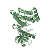
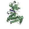
- PDB-1snu: CRYSTAL STRUCTURE OF THE UNPHOSPHORYLATED INTERLEUKIN-2 TYROSINE ... -
+ Open data

- Basic information
| Entry | Database: PDB / ID: 1snu | ||||||
|---|---|---|---|---|---|---|---|
| Title | CRYSTAL STRUCTURE OF THE UNPHOSPHORYLATED INTERLEUKIN-2 TYROSINE KINASE CATALYTIC DOMAIN | ||||||
 Components Components | Tyrosine-protein kinase ITK/TSK | ||||||
 Keywords Keywords | TRANSFERASE / PROTEIN KINASE / IMMUNOLOGY | ||||||
| Function / homology |  Function and homology information Function and homology informationNK T cell differentiation / gamma-delta T cell activation / Generation of second messenger molecules / cellular defense response / FCERI mediated Ca+2 mobilization / positive regulation of cytokine production / B cell receptor signaling pathway / T cell activation / non-specific protein-tyrosine kinase / non-membrane spanning protein tyrosine kinase activity ...NK T cell differentiation / gamma-delta T cell activation / Generation of second messenger molecules / cellular defense response / FCERI mediated Ca+2 mobilization / positive regulation of cytokine production / B cell receptor signaling pathway / T cell activation / non-specific protein-tyrosine kinase / non-membrane spanning protein tyrosine kinase activity / cell-cell junction / T cell receptor signaling pathway / adaptive immune response / intracellular signal transduction / signal transduction / zinc ion binding / ATP binding / nucleus / plasma membrane / cytosol Similarity search - Function | ||||||
| Biological species |  Homo sapiens (human) Homo sapiens (human) | ||||||
| Method |  X-RAY DIFFRACTION / SYNCHROTRON / MOLECULAR REPLACEMENT / Resolution: 2.5 Å X-RAY DIFFRACTION / SYNCHROTRON / MOLECULAR REPLACEMENT / Resolution: 2.5 Å | ||||||
 Authors Authors | Brown, K. / Long, J.M. / Vial, S.C. / Dedi, N. / Dunster, N.J. / Renwick, S.B. / Tanner, A.J. / Frantz, J.D. / Fleming, M.A. / Cheetham, G.M.T. | ||||||
 Citation Citation | Journal: J.Biol.Chem. / Year: 2004 Title: Crystal structures of interleukin-2 tyrosine kinase and their implications for the design of selective inhibitors. Authors: Brown, K. / Long, J.M. / Vial, S.C. / Dedi, N. / Dunster, N.J. / Renwick, S.B. / Tanner, A.J. / Frantz, J.D. / Fleming, M.A. / Cheetham, G.M. | ||||||
| History |
|
- Structure visualization
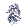
| Structure viewer | Molecule: MolmilJmol/JSmol |
|---|
- Downloads & links
-Download
| PDBx/mmCIF format | 1snu.cif.gz | 114.8 KB | Display | PDBx/mmCIF format |
|---|---|---|---|---|
| PDB format | pdb1snu.ent.gz | 88.9 KB | Display | PDB format |
| PDBx/mmJSON format | 1snu.json.gz | Tree view | PDBx/mmJSON format | |
| Others |  Other downloads Other downloads |
-Validation report
| Arichive directory | https://data.pdbj.org/pub/pdb/validation_reports/sn/1snuftp://data.pdbj.org/pub/pdb/validation_reports/sn/1snu | HTTPS FTP |
|---|
-Related structure data
| Related structure data | 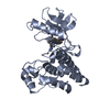 1sm2SC  1snxC S: Starting model for refinement C: citing same article ( |
|---|---|
| Similar structure data |


-Links
 PDBj
PDBj


- Assembly
| Deposited unit | 
| ||||||||
|---|---|---|---|---|---|---|---|---|---|
| 1 | 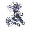
| ||||||||
| 2 | 
| ||||||||
| Unit cell |
|
-Components
| #1: Protein | Mass: 30104.424 Da / Num. of mol.: 2 / Fragment: CATALYTIC KINASE DOMAIN Source method: isolated from a genetically manipulated source Source: (gene. exp.) Homo sapiens (human) / Gene: ITK, LYK, EMT / Production host:  #2: Chemical | 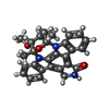  Mass: 466.531 Da / Num. of mol.: 2 / Source method: obtained synthetically / Formula: C28H26N4O3 / Comment: anticancer, antifungal, antibiotic, alkaloid*YM Mass: 466.531 Da / Num. of mol.: 2 / Source method: obtained synthetically / Formula: C28H26N4O3 / Comment: anticancer, antifungal, antibiotic, alkaloid*YM#3: Water | ChemComp-HOH / |  Mass: 18.015 Da / Num. of mol.: 197 / Source method: isolated from a natural source / Formula: H2O Mass: 18.015 Da / Num. of mol.: 197 / Source method: isolated from a natural source / Formula: H2O |
|---|
-Experimental details
-Experiment
| Experiment | Method: X-RAY DIFFRACTION / Number of used crystals: 1 |
|---|
- Sample preparation
| Crystal | Density Matthews: 3.01 Å3/Da / Density % sol: 59.16 % |
|---|---|
| Crystal grow | Temperature: 293 K / Method: vapor diffusion, hanging drop / pH: 5.7 Details: AMMONIUM SULFATE, SODIUM CITRATE, MAGNESIUM ACETATE, DTT, pH 5.7, VAPOR DIFFUSION, HANGING DROP, temperature 293K |
-Data collection
| Diffraction | Mean temperature: 100 K |
|---|---|
| Diffraction source | Source: SYNCHROTRON / Site: SRS  / Beamline: PX14.1 / Wavelength: 1.488 / Wavelength: 1.488 Å / Beamline: PX14.1 / Wavelength: 1.488 / Wavelength: 1.488 Å |
| Detector | Type: ADSC QUANTUM 4 / Detector: CCD / Date: Oct 28, 2003 |
| Radiation | Monochromator: Si 111 CHANNEL / Protocol: SINGLE WAVELENGTH / Monochromatic (M) / Laue (L): M / Scattering type: x-ray |
| Radiation wavelength | Wavelength: 1.488 Å / Relative weight: 1 |
| Reflection | Resolution: 2.5→20 Å / Num. all: 44498 / Num. obs: 22753 / % possible obs: 91.5 % / Observed criterion σ(F): 1 / Observed criterion σ(I): 1 / Redundancy: 2 % / Biso Wilson estimate: 50.7 Å2 / Rmerge(I) obs: 0.061 / Rsym value: 0.074 / Net I/σ(I): 11.3 |
| Reflection shell | Resolution: 2.5→2.64 Å / Redundancy: 1.6 % / Rmerge(I) obs: 0.296 / Mean I/σ(I) obs: 2.6 / Num. unique all: 2729 / Rsym value: 0.301 / % possible all: 76 |
- Processing
| Software |
| |||||||||||||||||||||||||
|---|---|---|---|---|---|---|---|---|---|---|---|---|---|---|---|---|---|---|---|---|---|---|---|---|---|---|
| Refinement | Method to determine structure: MOLECULAR REPLACEMENT Starting model: PDB ENTRY 1SM2 Resolution: 2.5→20 Å / Cross valid method: THROUGHOUT / σ(F): 3 / Stereochemistry target values: ENGH & HUBER
| |||||||||||||||||||||||||
| Refinement step | Cycle: LAST / Resolution: 2.5→20 Å
| |||||||||||||||||||||||||
| Refine LS restraints |
|
