 Movie
Movie Controller

[English] 日本語
 Yorodumi
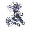
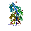
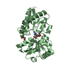
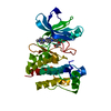
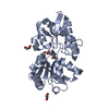
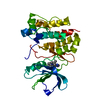
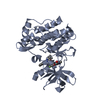
- PDB-4i84: The crystal structure of the Haemophilus influenzae HxuA secretio... -
+ Open data

- Basic information
| Entry | Database: PDB / ID: 4i84 | ||||||
|---|---|---|---|---|---|---|---|
| Title | The crystal structure of the Haemophilus influenzae HxuA secretion domain involved in the two-partner secretion pathway | ||||||
 Components Components | Heme/hemopexin-binding protein | ||||||
 Keywords Keywords | PROTEIN TRANSPORT / beta-helix / TWO-PARTNER SECRETION PATHWAY / HxuB | ||||||
| Function / homology |  Function and homology information Function and homology information | ||||||
| Biological species |  Haemophilus influenzae (bacteria) Haemophilus influenzae (bacteria) | ||||||
| Method |  X-RAY DIFFRACTION / SYNCHROTRON / MOLECULAR REPLACEMENT / Resolution: 1.5 Å X-RAY DIFFRACTION / SYNCHROTRON / MOLECULAR REPLACEMENT / Resolution: 1.5 Å | ||||||
 Authors Authors | Baelen, S. / Dewitte, F. / Clantin, B. / Villeret, V. | ||||||
 Citation Citation | Journal: Acta Crystallogr.,Sect.F / Year: 2013 Title: Structure of the secretion domain of HxuA from Haemophilus influenzae. Authors: Baelen, S. / Dewitte, F. / Clantin, B. / Villeret, V. | ||||||
| History |
|
- Structure visualization
| Structure viewer | Molecule: MolmilJmol/JSmol |
|---|
- Downloads & links
-Download
| PDBx/mmCIF format | 4i84.cif.gz | 247 KB | Display | PDBx/mmCIF format |
|---|---|---|---|---|
| PDB format | pdb4i84.ent.gz | 199.6 KB | Display | PDB format |
| PDBx/mmJSON format | 4i84.json.gz | Tree view | PDBx/mmJSON format | |
| Others |  Other downloads Other downloads |
-Validation report
| Arichive directory | https://data.pdbj.org/pub/pdb/validation_reports/i8/4i84ftp://data.pdbj.org/pub/pdb/validation_reports/i8/4i84 | HTTPS FTP |
|---|
-Related structure data
| Related structure data |  2odlS S: Starting model for refinement |
|---|---|
| Similar structure data |


-Links
 PDBj
PDBj- Assembly
| Deposited unit | 
| ||||||||
|---|---|---|---|---|---|---|---|---|---|
| 1 | 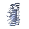
| ||||||||
| 2 | 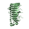
| ||||||||
| Unit cell |
|
-Components
| #1: Protein | Mass: 32967.695 Da / Num. of mol.: 2 / Fragment: UNP residues 24-324 Source method: isolated from a genetically manipulated source Source: (gene. exp.) Haemophilus influenzae (bacteria) / Strain: ATCC 51907 / DSM 11121 / KW20 / Rd / Gene: HI_0264, hxuA, KW20 / Production host: #2: Water | ChemComp-HOH / |  Mass: 18.015 Da / Num. of mol.: 429 / Source method: isolated from a natural source / Formula: H2O Mass: 18.015 Da / Num. of mol.: 429 / Source method: isolated from a natural source / Formula: H2O |
|---|
-Experimental details
-Experiment
| Experiment | Method: X-RAY DIFFRACTION / Number of used crystals: 1 |
|---|
- Sample preparation
| Crystal | Density Matthews: 2.18 Å3/Da / Density % sol: 43.5 % |
|---|---|
| Crystal grow | Temperature: 293 K / Method: vapor diffusion, hanging drop / pH: 6.5 Details: 0.095M TRI SODIUM CITRATE pH 5.6, 5% GLYCEROL, 19% ISOPROPANOL, 12% PEG4000, 0.5M IMIDAZOLE pH 6.5, VAPOR DIFFUSION, HANGING DROP, temperature 293K |
-Data collection
| Diffraction | Mean temperature: 100 K |
|---|---|
| Diffraction source | Source: SYNCHROTRON / Site: SOLEIL  / Beamline: PROXIMA 1 / Wavelength: 0.98011 Å / Beamline: PROXIMA 1 / Wavelength: 0.98011 Å |
| Detector | Type: ADSC QUANTUM 315r / Detector: CCD / Date: Sep 29, 2011 |
| Radiation | Monochromator: CHANNEL CUT CRYOGENICALLY COOLED MONOCHROMATOR CRYSTAL Protocol: SINGLE WAVELENGTH / Monochromatic (M) / Laue (L): M / Scattering type: x-ray |
| Radiation wavelength | Wavelength: 0.98011 Å / Relative weight: 1 |
| Reflection | Resolution: 1.5→41.9 Å / Num. all: 89861 / Num. obs: 89775 / Observed criterion σ(F): 2 / Observed criterion σ(I): 2 |
| Reflection shell | Resolution: 1.5→1.59 Å |
- Processing
| Software |
| ||||||||||||||||||||||||||||||||||||||||||||||||||||||||||||||||||||||
|---|---|---|---|---|---|---|---|---|---|---|---|---|---|---|---|---|---|---|---|---|---|---|---|---|---|---|---|---|---|---|---|---|---|---|---|---|---|---|---|---|---|---|---|---|---|---|---|---|---|---|---|---|---|---|---|---|---|---|---|---|---|---|---|---|---|---|---|---|---|---|---|
| Refinement | Method to determine structure: MOLECULAR REPLACEMENT Starting model: PDB ENTRY 2ODL Resolution: 1.5→41.9 Å / Cor.coef. Fo:Fc: 0.971 / Cor.coef. Fo:Fc free: 0.955 / SU B: 2.648 / SU ML: 0.045 / Cross valid method: THROUGHOUT / σ(F): 2 / ESU R: 0.083 / ESU R Free: 0.077 / Stereochemistry target values: MAXIMUM LIKELIHOOD / Details: HYDROGENS HAVE BEEN ADDED IN THE RIDING POSITIONS
| ||||||||||||||||||||||||||||||||||||||||||||||||||||||||||||||||||||||
| Solvent computation | Ion probe radii: 0.8 Å / Shrinkage radii: 0.8 Å / VDW probe radii: 1.4 Å / Solvent model: MASK | ||||||||||||||||||||||||||||||||||||||||||||||||||||||||||||||||||||||
| Displacement parameters | Biso mean: 20.866 Å2
| ||||||||||||||||||||||||||||||||||||||||||||||||||||||||||||||||||||||
| Refinement step | Cycle: LAST / Resolution: 1.5→41.9 Å
| ||||||||||||||||||||||||||||||||||||||||||||||||||||||||||||||||||||||
| Refine LS restraints |
| ||||||||||||||||||||||||||||||||||||||||||||||||||||||||||||||||||||||
| LS refinement shell | Resolution: 1.5→1.538 Å / Total num. of bins used: 20
|
